The INM endeavors to pursue captivating research projects within a rich and highly heterogeneous environment. The tutorials offered during our retreat have traditionally presented an exceptional opportunity to embrace this diversity and expand one’s understanding of scientific concepts, methodological approaches, and future challenges that extend beyond our daily work routine. In the INM Retreat 2024, we are pleased to announce that researchers from various institutes once again extend their offer to share their knowledge and expertise on selected topics.
Time: Tuesday – November 19, 9:00 – 13:00
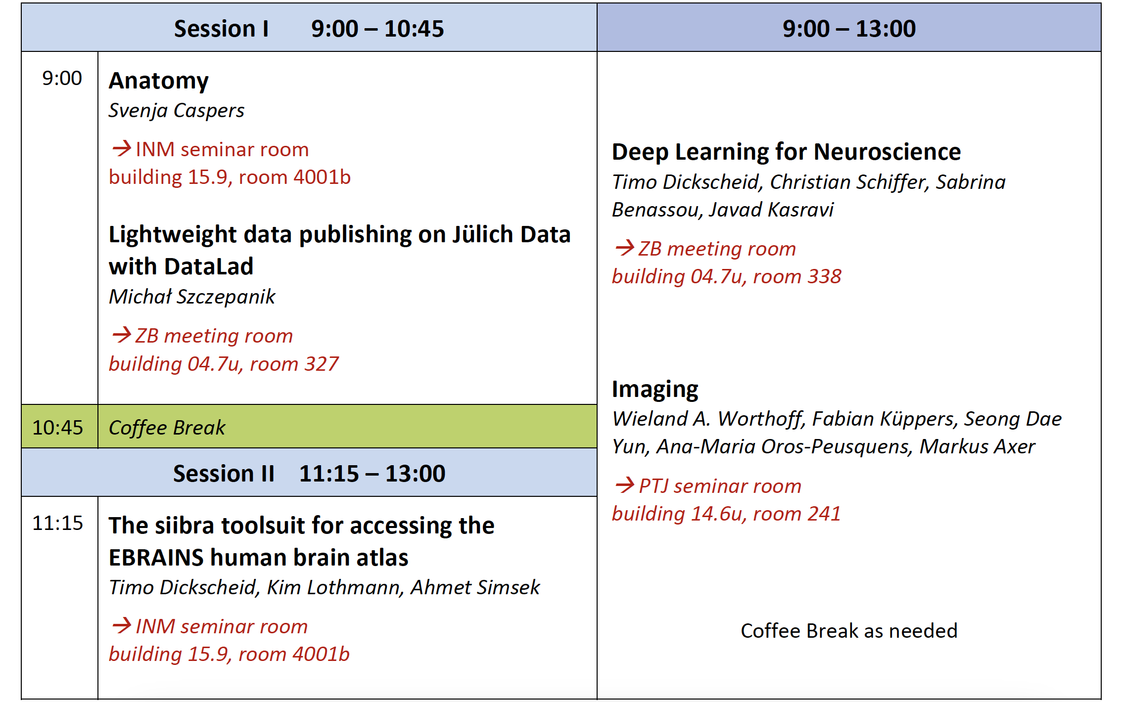
Anatomy
9:00 – 10:45 | INM seminar room | building 15.9, room 4001b
Svenja Caspers
The tutorial will cover the basic principles of neuroanatomy, including major landmarks for orientation and understanding of basic functions. It will combine insights about the brain’s structure across scales and organizational levels. Accompanying the theoretical foundations, the tutorial will provide hands-on training on live dissection of brain sections.
Lightweight data publishing on Jülich Data with DataLad
9:00 – 10:45 | ZB meeting room | building 04.7u, room 327
Michał Szczepanik
Jülich DATA (data.fz-juelich.de) is the central institutional repository for research data of the research center Jülich and supports sharing, preserving, citing, exploring, and analyzing research data with descriptive metadata, without hosting large files. In this tutorial, participants will discover how DataLad (datalad.org) can integrate with Dataverse and have the best of both worlds: Discoverability and metadata with Jülich DATA, and actionable data tracking with DataLad. With conceptual and hands-on elements, we will learn how to publish or clone lightweight DataLad datasets to and from Jülich DATA.
Deep Learning for Neuroscience
9:00 – 13:00 | ZB meeting room | building 04.7u, room 338
Timo Dickscheid, Christian Schiffer, Sabrina Benassou, Javad Kasravi
Machine Learning – in particular deep learning – has become an indispensable tool for analyzing large neuroscience datasets. The Helmholtz AI team at Jülich is closely connected to these developments and supports research activities at the intersection of AI, high-performance computing (HPC) and neuroscience. Many of the methods and solutions are not limited to neuroscience and medical applications, but can be transferred to different tasks and scientific domains.
This tutorial we will give an overview of state-of-the-art deep learning methods in the context of biomedical image analysis and show concrete examples in INM where deep learning already supports neuroscientists in analyzing their data.
The second part of this tutorial will offer a hands-on course on how to bring deep learning pipelines on JSC’s HPC systems.
If you plan to join the hands-on part, please read the prerequisites here carefully.
Imaging
9:00 – 13:00 | PTJ seminar room | building 14.6u, room 241
|
09:00 |
Session I – MR Imaging 09:00 – 09:45 – Introduction to MRI (Dr. Wieland A. Worthoff) 09:45 – 10:00 – (questions) 10:00 – 10:45 – Insights into brain function: Advances in functional MRI methods (Dr. Fabian Küppers and Dr. Seong Dae Yun) |
|
10:45 |
Coffee Break (with possibility to interact with the speakers) |
|
11:15 |
Session II – Imaging Fibers 11:15 – 11:45 – Diffusion Imaging (Dr. Ana-Maria Oros-Peusquens) 12:00 – 12:45 – 3D-polarized light imaging (3D-PLI) (Prof. Dr. Markus Axer) |
The tutorial will provide insights into advanced imaging techniques, focusing on basics of MRI, functional MRI (fMRI), Diffusion Tensor Imaging (DTI), and 3D-Polarized Light Imaging (3D-PLI). The tutorial is designed to explore imaging techniques used for understanding both brain structure and function, emphasizing the latest advances in the field. Participants will gain foundational knowledge of each method, followed by contents related to applications in neuroscience research and clinical practice.
Session I – MR Imaging
- Introduction to MRI (09:00 – 09:45)
This session introduces the fundamental principles of Magnetic Resonance Imaging (MRI), beginning with spin physics to establish a solid understanding of MRI functionality. It will also briefly discuss the advantages and disadvantages of ultra-high field strength systems (7 Tesla and above).
- Insights into Brain Function: Advances in Functional MRI Methods (10:00 – 10:45)
This tutorial begins by explaining the fundamental principles behind fMRI and how it measures changes in blood oxygenation to infer neural activity. This session will explore the advances in functional MRI (fMRI) methods used to understand brain activity. It will cover recent developments in the techniques and protocols used for functional imaging, such as improvements in signal acquisition and noise reduction. A special focus will be placed on advanced applications like resting-state fMRI and task-based fMRI, providing a deeper understanding of how these imaging techniques contribute to our knowledge of brain function.
Coffee Break (10:45 – 11:15)
Participants will have the chance to interact informally with the speakers over coffee, encouraging networking and discussions about the topics presented.
Session II – Imaging Fibers
- Diffusion Imaging (11:15 – 11:45)
This tutorial will provide an introduction to diffusion and Diffusion Tensor Imaging (DTI), a powerful MRI-based technique for mapping the fibre structure and structural connectivity of the brain. Participants will learn about the principles of water diffusion and how DTI can be used to create detailed images of the brain's fibre tracts. Additionally, we will exemplify clinical and research applications of DTI, particularly in assisting brain tumour surgery and assessing the structural connectivity of the brain. - 3D-Polarized Light Imaging (3D-PLI) (12:00 – 12:45)
This session will introduce 3D-Polarized Light Imaging (3D-PLI), a technique used for visualizing the orientation of nerve fibers in the brain. 3D-PLI provides a unique approach to studying the organization of the brain's white matter, complementing traditional imaging methods like DTI. Participants will learn about the principles behind 3D-PLI, its applications in neuroscience research, and how it contributes to our understanding of the intricate structural organization of neural networks.
The siibra toolsuit for accessing the EBRAINS human brain atlas
11:15 – 13:00 | INM seminar room | building 15.9, room 4001b
Timo Dickscheid, Kim Lothmann, Ahmet Simsek
siibra is a software tool suite implementing an openly accessible brain atlas framework which connects multimodal datasets from different resources to anatomical structures in reference spaces at different spatial scales. The tool suite is designed to address both interactive exploration through an interactive 3D web viewer (siibra-explorer) as well as integration into data analysis and simulation workflows with a comprehensive Python library (siibra-python). In this session, we first introduce the multidimensional concept of the atlas framework and explore some key features such as the BigBrain interactively. We then turn to concrete programming tutorials in Python. These include fetching brain region maps, accessing the BigBrain dataset, and extracting multimodal regional features such as cortical thicknesses, cell and neurotransmitter densities, gene expressions and connectivity data. We will finish with some concrete data analysis examples.
