Choose timezone
Your profile timezone:
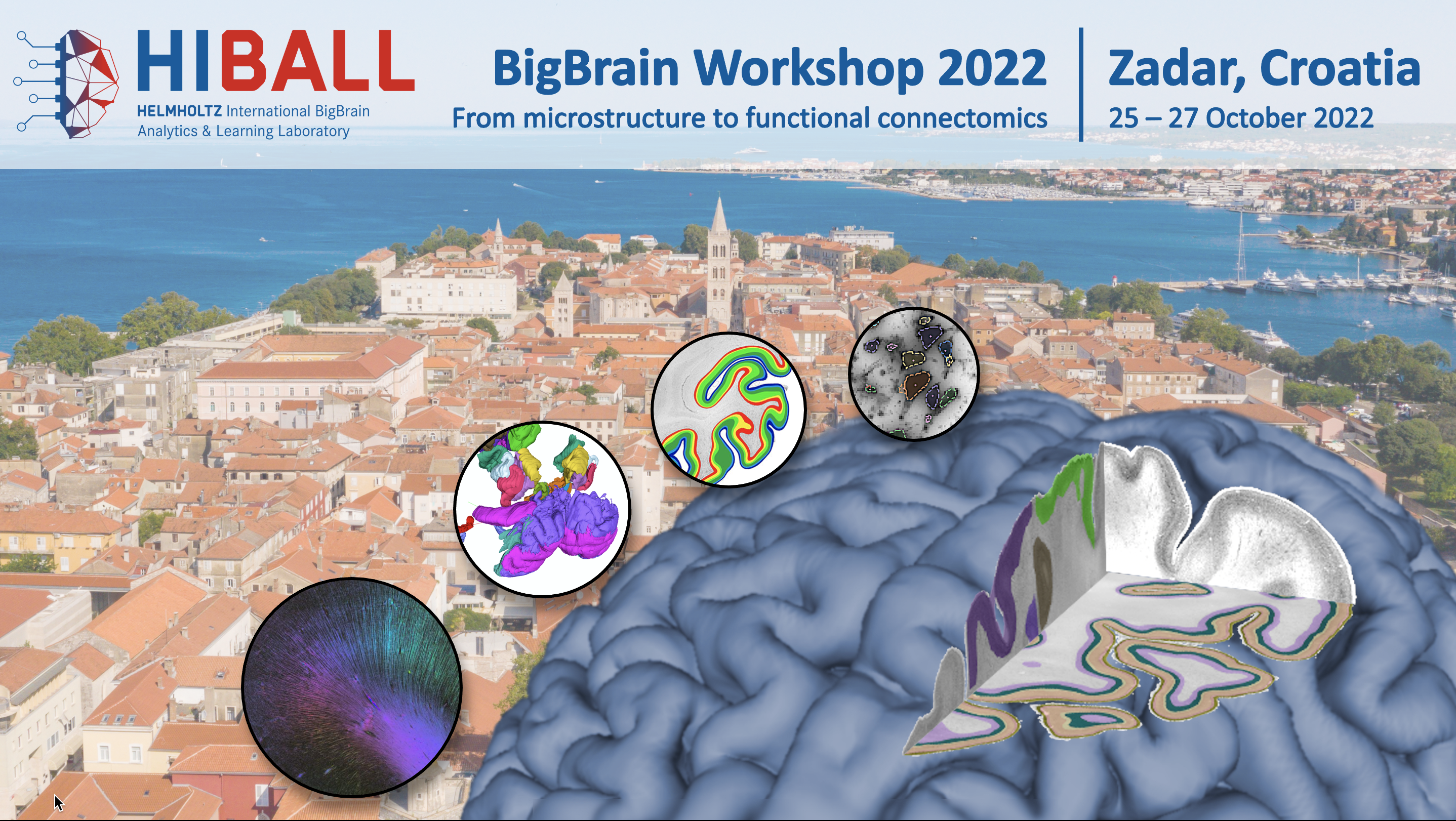
We are pleased to announce the 6th BigBrain Workshop as an in-person event taking place on
October 25-27, 2022 in Zadar, Croatia.
With this workshop we reach out to the international community of BigBrain users and invite researchers from our global network (and beyond) to present their work and to discuss future prospects of the BigBrain data and tools, in particular to discuss how to better leverage high performance computing (HPC) and artificial intelligence (AI) to create multimodal, multiresolution tools for the high resolution BigBrain and related datasets. We are proud to announce Mu-ming Poo and Ivica Kostović as our keynote speakers.
The BigBrain Workshop will be organized as a symposium, with both invited speakers and contributed talks as well as a poster and demo session. We welcome short abstracts of current work and/or short proposals for future initiatives related to the BigBrain or similar data.
As an HBP partnering project, HIBALL is committed to HBP’s training activities and supports this with joint events and participation in the curriculum, through the work of our shared members, offering young researchers a great opportunity to showcase their research work and practise their mentoring skills.
We are pleased to announce already that the BigBrain Workshop will be held in conjunction with the Autumn HBP Young Researchers Event, taking place as a full day event on October 25, on-site at the conference venue. A separate registration is required!
The event is free of charge but prior registration is required.
We provide a limited contingent of rooms at the conference venue, as well as information about further accommodation.
Important dates
Organizing Committee
|
Montreal Neurological Institute |
Institute of Neuroscience and Medicine (INM-1) |
|
|
Please contact the program committee if you have any questions. We will continuously update the information on this page and also share information via Twitter (@BigBrainProject) and e-mail.
 |
 |
||
 |
 |
 |
|
For some registered participants, mails from this platform seem to end up as spam. To make sure to receive all information, please check your junk folder.
Per default all times are given in CEST. To make sure to refer to your local time, please set your time zone accordingly. Click the menu at the top right of this page.
 |
www.bigbrainproject.org |  |
@BigBrainProject |
Katrin Amunts and Alan Evans
Transcriptome, Connectome and Neuromodulation of the Primate Brain
Mu-Ming Poo
Institute of Neuroscience and Center for Excellence in Brain Science and Intelligence Technology, Chinese Academy of Sciences
Future trends in microstructural connectivity
Panel:
Prof MuMing Poo, Institute of Neuroscience, Chinese Academy of Sciences; Shanghai Center for Brain Science and Brain-inspired Technology
Prof Ivica Kostovic, Croatian Institute for Brain Research
Prof Boris Bernhardt, The Neuro, McGill University
Prof Amir Shmuel, The Neuro, McGill University
Prof Dr Hanchuan Peng, Allen Institute for Brain Science
Moderation: Alan Evans and Katrin Amuts
The cerebral cortex is made up of roughly six horizontally organized layers with unique cytoarchitectural properties distributed across the cortical mantle. This gradual variation of laminar structure is a fundamental principle of cortical organization. The similarity of regions in their laminar structure, with roots in development, is suggested to relate to the likelihood, strength and direction of their connectivity. Current accounts of laminar structure variation are mainly based on theory-driven approaches in which histological samples of the cortex are labeled into discrete types based on visual inspection of laminar features. Here, leveraging on a data-driven map of the six cortical layers in the BigBrain, we aimed to quantitatively characterize the gradual variation of laminar structure in the cortex. We identified an organizational axis of laminar thickness covariance which differentiated the dominance of infragranular and supragranular layer thickness and in general followed a rostro-caudal trajectory. This axis was co-aligned with the cortical hierarchy such that infragranular-dominant regions towards the rostral pole of the cortex were overall higher up this functional hierarchy. Furthermore, laminar thickness variation was linked to connectivity, with regions with similar laminar thickness showed higher likelihood and strength of connectivity. Laminar thickness covariance was also related to structural covariance, reflecting shared developmental/maturational and genetics effects of the regions with similar laminar structure, which hints at developmental origins of laminar structure variability. In sum, we describe the organization of layer-wise thickness covariation in the cortical mantle and how it relates to structure and function.
Introduction One important feature of the human brain is structural asymmetry in the homologous contralateral anatomical regions. Left-right asymmetries of cortical thickness and surface area exist widely across the cortical mantle but intracortical microstructure asymmetry is rarely studied. Here we evaluate the asymmetry of microstructural organization in the human cortex combining post mortem and in vivo datasets.
Methods We assessed asymmetry of microstructural intensity using a 20-μm ultra-high–resolution 3D histological reconstruction (cell-body staining) of a post mortem brain (BigBrain, male donor, age = 65 years), together with in vivo myelin-sensitive magnetic resonance imaging (MRI) data including young adult Human Connectome Project (HCP, n=1101) T1w/T2w maps and Microstructure-Informed Connectomics (MICA-MICs, n=50) quantitative T1 (qT1) relaxometry. We downsampled the cortical maps using a multi-modal parcellation and Cole-Anticevic network atlas. Asymmetry of intracortical organization was computed by using microstructure profile covariance (MPC) gradients of intracortical equivolumetric surfaces.
Results We observed layer-specific cytoarchitectural asymmetry of the human cortex using post mortem data (Figure 1A). All p-values displayed are less than 0.001. Layer IV showed highest spatial similarity between layers, with mean r = 0.739. We also observed increased rightward asymmetry with deeper intracortical depth in dorsal attention network (DAN, r = -0.679), and increased leftward asymmetry with deeper intracortical depth in networks including somatomotor (SMN), cingulate-opercular (CON, r = 0.652), language (Lan., r = 0.620), and posterior-multimodal (PMN, r = 0.856). Following, we evaluated asymmetry of intracortical profile covariance. The principal gradient of MPC (MPC G1) followed an anterior-position direction. Along this axis, visual cortices showed rightward asymmetries but posterior middle temporal gyrus and inferior parietal cortices showed leftward asymmetries (Figure 1B). Then, we evaluated whether similar patterns of asymmetry could be observed in vivo, using intra-cortical microstructural proxies. Visual and somatomotor cortices showed high myelination intensity for both qT1 and T1w/T2w data (Figure 1C). The left-right asymmetry of intensity followed an anterior-posterior direction (Figure 1D), as well as the asymmetry of MPC G1 (Figure 1E). T1w/T2w MPC G1 showed strongest leftward asymmetry in orbito-affective (OAN, Cohen’s d intra-hemisphere = 0.465, Cohen’s d inter-hemisphere = 0.478), frontoparietal (FPN, Cohen’s d intra-hemisphere = 1.314, Cohen’s d inter-hemisphere = 1.232), and language regions (Cohen’s d intra-hemisphere = 0.447, Cohen’s d inter-hemisphere = 0.414). Strongest rightward asymmetry was observed in PMN (Cohen’s d intra-hemisphere = -0.357, Cohen’s d inter-hemisphere = -0.265), SMN (Cohen’s d intra-hemisphere = -0.353, Cohen’s d inter-hemisphere = -0.769), and second visual network (Vis2, Cohen’s d intra-hemisphere = -0.600, Cohen’s d inter-hemisphere = -0.447). The spatial pattern is similar between T1w/T2w and qT1 (r intra-hemisphere = 0.556 and r inter-hemisphere = 0.542).
Conclusions In sum, we find that the cytoarchitectural asymmetry of the cortex differs between laminar structures, with intracortical changes in language, somatomotor, posterior multimodal, and dorsal attention networks, in particular in posterior cortical regions. Regarding the in vivo maps of intracortical microstructural asymmetry, our two measures suggest similar results in microstrural intensity but subtle differences in organization features.
Background. The claustrum is a “sheet-like” telencephalic grey matter structure, wrapped by the capsulae extrema and externa, and tucked between the putamen and insula. Anatomical studies have revealed that the claustrum is one of the most connected of any brain structure (by volume), and demonstrates reciprocal connections across the entire cortical mantle. Though this remarkable connectivity has given rise to a gamut of hypotheses, very little is known about claustral function due to its complex shape, thinness and location, hampering in vivo investigation with conventional MRI. Specifically, typical imaging resolution at 3 Tesla is insufficient to differentiate the claustrum from the insula and putamen, and all but precluded automated segmentation methods.
Objective. We are seeking to clarify claustral structure and function using MRI at ultra-high field, with a spatial resolution sufficient, in principle, to disambiguate the claustrum from its surrounding structures. However, these efforts have been limited by the lack of a ‘ground truth’ claustral reference. Our objective is to provide this reference by segmenting the claustrum in the BigBrain dataset. By making our segmentation publicly available, we will provide a shared benchmark to quantify the promise, and pitfalls, of claustral investigation at ultra-high fields.
Methods. We have segmented the right claustrum on the 100 micron isotropic BigBrain in MNI ICBM-152 space. We found that ITK-SNAP’s semi-automatic segmentation methods gave rise to both false positives (inclusion) and negatives (exclusion), so opted to delineate the structure manually. Our manual segmentation was informed by simultaneous visualization on all three planes; in cases of ambiguous inclusion/exclusion, we consistently deferred to the axial plane. To quantify the integrity of claustral reconstruction at ultra-high field, we followed the same manual segmentation procedure using a 0.7mm isotropic MP2RAGE dataset on healthy human subjects (N=6) collected on a Siemens 7 Tesla at Maastricht University, Netherlands.
Results. The unparallelled resolution afforded by BigBrain has underscored the claustrum’s challenging anatomy. Generally, our reference segmentation is consonant with claustral descriptions in histological literature, though one unanticipated finding is that the right claustrum appears larger than reported (spanning approximately 40mm rostrocaudally and 25mm dorsoventrally, vs. 38mm and 22mm, respectively). Our comparative evaluation of the claustrum in the Maastricht dataset suggests that the claustrum is only partially captured at 7 Tesla; notably, its extreme extents evade detection. We are presently working to quantify the spatial overlap between the BigBrain reference and Maastricht datasets, and estimate heterogeneity between participants in the latter (results forthcoming for presentation).
Discussion. We anticipate that our ‘ground truth’ BigBrain segmentation will be of interest to researchers in its own right. Further, we anticipate our work will delineate exactly how (native) ultra-high field MRI falls short of adequately capturing the claustrum in vivo. We hope that our BigBrain reference may inform guided segmentation efforts, which we believe may be necessary for claustral research at ultra-high field. Excitingly, the clastrum’s span appears sufficient to seed connectivity, opening the prospect of moving from microstructure to functional connectomics, and in so doing, unraveling enduring mysteries of claustral function.
Introduction Buried in the temporal lobes, the amygdala is a crucial structure for emotion and social cognition. Detailed post mortem studies have highlighted several subdivisions within the amygdala, each with distinct cytoarchitectural characteristics and distinguishable connectivity profiles to other regions of the brain (Kedo et al., 2017). However, these atlases rely on labour-intensive visual inspections of histological specimens performed by expert neuroanatomists. Here, we build a multiscale framework grounded in foundational histological studies of the neocortex to map amygdala cytoarchitecture in a data-driven manner. We cross-reference this approach against manually segmented labels provided in established atlases of amygdala anatomy(Amunts et al., 2020).
Methods. Histological data of the amygdala was obtained from the 100μm BigBrain dataset (Amunts et al., 2013) (Fig1A), in which image intensity values provide direct measurements of brain cytoarchitecture (soma size and density). The amygdalae were isolated using an existing manual segmentation of subcortical structures (Xiao et al., 2019). This segmentation was warped to BigBrain histological space using co-registration strategies aggregated in the BigBrainWarp toolbox (Paquola et al., 2021). Guided by prior neuroanatomical studies of cortical histology (Palomero-Gallagher Zilles, 2018), we built a histological feature bank of the amygdala. Specifically, feature selection leveraged the parameterization of central moments mapping intensity variations across the amygdala. We used a radiomics approach (van Griethuysen et al., 2017) to compute voxel-based maps for each of the selected first-order features (mean, variance, skewness, and kurtosis) at 5 different kernel sizes. This resulted in 20 distinct feature maps, reflecting variations in intensity distributions within the amygdala from finer to coarser scales (Fig1A). To capture and visualize the underlying structure of amygdala cytoarchitecture, we applied UMAP, a non-linear dimensionality reduction technique (McInnes et al., 2018), to our microstructural feature bank (Fig1B). We then leveraged openly available probabilistic maps of amygdala subnuclei (1,8,9) labelling each voxel to its highest probability subdivision (Fig1C). For further neuroanatomical contextualization, correlations were calculated between voxel-wise UMAP components values and corresponding spatial coordinates (Fig1C). These steps were then repeated with 7T quantitative T1 (qT1) images of 6 subjects (Fig1D).
Results. Our data-driven process could partly recover ground-truth anatomical subdivisions of the amygdala. Notably, the three anatomical subdivisions seemed to primarily follow the direction of U2 (Fig1C). Furthermore, U1 seemed to primarily vary on the medial-lateral axis (r=0.3186), whereas U2 was primarily and highly correlated to the inferior-superior axis (r=0.88), both correlations were statistically significant with a variogram matching test. UMAP-driven visualizations of the histological feature space suggest our dimensionality reduction approach could capture global intensity covariations across moments. When reproducing the analyses on qT1 images, the UMAP components illustrated similar trends from their respective feature banks (Fig1D). U2 generally captures more inferior-superior gradients and U1 more medial-lateral variance. Statistical analysis of these similarities is currently being investigated.
Conclusions. We propose data-driven approach for investigations of amygdala cytoarchitecture. This novel method, together with the reproducible findings found in qT1 images, shows great potential for an efficient and accurate representations of cytoarchitecture that can support investigations of subject-specific structure-function coupling in subcortical structures.
The hippocampus (or archicortex) has a complex folded laminar structure that can be fully appreciated in the multiplanar views afforded by 3D BigBrain [1]. Here, we will look at how analysis of the hippocampus and hippocampal subfields can be performed using the same principles as surface-based analysis of the neocortex and neocortical parcels [2]. That is, at high resolution the various folds or ‘digitations' within the hippocampus resemble neocortical gyrifications. By carefully fitting a surface to these folds, we extract depth-wise profiles of neuronal distributions across the 3D BigBrain hippocampi. Two complementary sets of results show that: i) within the hippocampus, unsupervised clustering of these profiles yields boundaries that almost perfectly overlap with the classical definitions of hippocampal subfields [3], and, ii) between the hippocampus and neocortex, there exists a continuum or gradient of changes in profile similarity, with greatest complexity in the neocortex [4].
In the domain of MRI, spatial resolution is considerably lower but with strong prior information a surface can accurately fit the folds of the hippocampus along its natural anterior-posterior and medial-lateral 2D axes [5]. Using MRI data from the Human Connectome Project dataset [6], we show that structural measures track the hippocampus’ medial-lateral axis, whereas resting state connectivity (rsfMRI) tracks its anterior-posterior axis. That is, examining structural measures like thickness, gyrification, and curvature, there are clear differences across the medial-lateral extent of the hippocampus in line with the hippocampal subfields. However, the primary differences in functional connectivity are aligned along the anterior-posterior axis of the hippocampus in a geodesic fashion. That is, the anterior hippocampus tends to show greater connectivity with anterior temporal and inferior frontal regions of the neocortex, whereas the posterior hippocampus tends to show greater connectivity to the anterior cingulate, medial visual, temporal-parietal junction, and lateral frontal neocortex.
Together these results present a simple model for understanding the hippocampus: it shows a stereotyped microstructure across its medial-lateral axis that repeats across its anterior-posterior axis with different input/output connectivity to the rest of the brain. This organizing principle is obviated by treating the hippocampus as a 2D folded structure with two natural geodesic axes rather than as a subcortical volume with subfields as subvolumes, as in many previous works.
References
1. Amunts K, et al. BigBrain: an ultrahigh-resolution 3D human brain model. Science. 2013;340: 1472–1475.
2. DeKraker J, et al. Surface-based hippocampal subfield segmentation. Trends Neurosci. 2021;44: 856–863.
3. DeKraker J, et al. Hippocampal subfields revealed through unfolding and unsupervised clustering of laminar and morphological features in 3D BigBrain. Neuroimage. 2020;206: 116328.
4. Paquola C, et al. Convergence of cortical types and functional motifs in the human mesiotemporal lobe. Elife. 2020;9. doi:10.7554/eLife.60673
5. DeKraker J, et al. Unfolding the hippocampus: An intrinsic coordinate system for subfield segmentations and quantitative mapping. Neuroimage. 2018;167: 408–418.
6. Van Essen DC, et al. The WU-Minn Human Connectome Project: an overview. Neuroimage. 2013;80: 62–79
Introduction. We are developing receptor architectonic rat brain maps to expand the Waxholm Space Atlases of the Rat Brain with functionally relevant data. This study focused on the development of 3D maps of the whole rat iso and proisocortex, regarding the heterogeneous distribution of glutamatergic (AMPA, kainate, NMDA), serotoninergic (5 HT1A, 5 HT2), adrenergic α1 and muscarinic (M2, M3) receptors and their correlation with cytoarchitectonic borders.
Methods. Alternating coronal serial sections (thickness 20 µm) through entire rat brains are used for silver cell-body staining and quantitative in vitro autoradiographic labeling of eight receptor types to identify cortical borders based on cyto and receptor architectonic heterogeneities, respectively. Mean receptor densities were measured in each identified area, and the ensuing parcellation brought into the Waxholm Rat Atlas stereotaxic space1.
Results and Conclusion. Over 50 distinct areas were identified in the rat iso- and proisocortex based on multimodal analysis of the regional and laminar differences in cytoarchitecture and eight different receptors. The isocortex encompasses frontal areas Fr1-Fr3, FrHL, FrFL; parietal areas Par1, Par2, ParHL and ParFL, ventral and posterior parietal areas (ParVr, ParVc and ParPd, ParPv, respectively); granular insular areas GIa, GIp; temporal areas Te1, Te2d, Te2v, Te3r and Te3v; occipital areas Oc1B, OC1M, medial areas Oc2MM and Oc2ML and lateral areas Oc2Lr, Oc2Lc, Oc2Lid and Oc2Liv; and finally, ectorhinal areas EctD and EctV. Within the proisocortex we identified orbitofrontal areas DLO, LO, VLO, VO and MO; cingulate areas Cg1, Cg1’, Cg2d, Cg2d’, Cg2v, Cg2v’, Cg3; agranular retrosplenial area RSA; dysgranular insular DI; posterior ectorhinal EctP and dorsal and ventral perirhinal areas PRhD and PRhV. As described in primates, rat primary sensory areas represented different receptor distribution patterns e.g., higher M2 and 5 HT2 receptor densities in layer IV, which is the main target of sensory input. Contrarily, kainate and α1 receptors showed lower densities in layer IV. Additionally. to the frontal areas Fr1 and Fr2, FrHL and FrFL were marked by higher mean densities of the eight investigated receptor types. Whereas the heterogenous laminar distributions of kainate, M2, M3 and 5 HT2 receptors represented the borders to the adjacent areas ParHL, ParFL and further the whole parietal cortex. Differences (e.g., AMPA), as well as homologies (e.g., NMDA) in receptor architecture were analyzed within the rat cingulate and retrosplenial areas, compared to primates.
The ensuing map can be used for cyto and receptor architectonically informed analyses of future in vivo studies via the Waxholm Rat Atlas stereotaxic space3, and thus constitutes a valuable resource for the neuroscientific community.
Reconstruction of a BigBrain dataset from serial histological sections is highly labour and time intensive and requires a significant technical effort. To obtain a high-quality 3D reconstruction from accurate image registration, it is necessary to correct images from artifacts that can arise from cutting and from further histological processing steps (e.g., cracks, ripples, holes), even if all processing steps are performed with great care and expertise. Previous work on such a dataset showed that the effort for repair is the most critical factor in the total time required to reconstruct the dataset. Strategies for these repairs are presented, discussed, and classified in terms of their effectiveness, amount of required manual work and computational requirements. An overview of typical damages seen in histological sections is provided with an estimate of their relative frequency. Due to the number and variety of these damages, fully automatic procedures quickly prove to be insufficient. To enable time-effective repair of thousands of sections, optimized, and easy-to-use frameworks as well as quality control (QC) and consistent provenance tracking tools are essential. Methods that have already been applied in practice are presented.
We developed a semi-automatic procedure considering different approaches depending on the overall quality of individual sections. E.g., if the damage is present in an individual section, information from adjacent sections can be used for an automatic repair, which is not feasible if the damage affects several consecutive sections. Further, damages should not be repaired if they were already present after removal of the brain, because such damages are also imaged in the MR dataset, used hereby as an undistorted shape reference in the reconstruction, and their elimination could mislead the registration. Our processing pipeline is based on a recursive multi-resolution approach with integrated provenance tracking, initially generating a coarse-resolution reconstruction that is increased in iterative steps to the final resolution of 0.02x0.02x0.02 mm.
Based on this general framework, we will report on the progress in the repair of the so-called BigBrain2, which consists of 7676 histological sections. Every 5th section was fully repaired manually. Subsequently, a semi-automatic repair framework was applied to the intermediate sections. Each original, unrepaired image was first nonlinearly transformed to the immediately adjacent repaired sections. Using the computed transformation, the repaired image was then transformed into the space of the adjacent unrepaired sections and used to assist the automatic repair of the section. In contrast to our previous approaches, the protocolled repair information is additionally used to improve the repair of damages affecting several adjacent sections. To evaluate the results, an easy-to-use online tool for quick visual control of the sections in the different processing stages has been developed. In cases QC indicates that the result does not meet the required quality standards, the section is returned into the pipeline for another iteration.
Our intermediate results show that the improved approach for semi-automatic repairs with provenance tracking and online QC accelerates the very time-critical repair process within the overall 3D BigBrain dataset reconstruction.
Blind image restoration aims at recovering high-quality images from data deteriorated by artifacts due to noise, low resolution, motion blur, lossy compression or other, in the absence of any knowledge about the degradation function.
In normal settings, restoration relies on pairing an input degraded image with a reference image in good condition to provide prior information for building the restoration model.
However, the lack of access to high quality reference priors limits the practical applicability of blind restoration models, while the limited capacity of learning dictionaries restricts the diversity and richness of restored details.
We propose a blind super-resolution model to address the resolution upscaling scenario when the function for mapping high-resolution and low-resolution images is unknown.
Our solution relies on three training modules with different learning objectives: 1. a degradation-aware network (U-Net) to synthesize the high resolution image, given a low resolution image and the corresponding blur kernel; 2. a pre-trained generative adversarial network (GAN) to be used as prior, bridged to the U-Net by a latent code mapping and several channel-split spatial feature transforms (CS-SFTs); and 3. a rational polynomial image interpolation into deep convolutional neural networks (CNNs) to retain details.
The U-Net is designed to remove complex degradation and extract two kinds of features: i) latent features to map the input image to the closest latent code in the GAN, and ii) multi-resolution spatial features for modulating the GAN features which increase the receptive field for large blur elimination. Part of the GAN features that contribute to image fidelity are spatially modulated by the CS-SFTs, while left-out features that contribute to image realness directly pass through (Figure, top left). These features are subsequently mapped to the intermediate latent codes for improved preservation of the semantic properties. The latent codes are then passed through each convolution layer in the pre-trained GAN, and generate (GAN) features for each resolution scale. The model is trained on synthetic data produced using a known degradation model that approximates realistic low-quality images, and generalize to real images during inference.
We evaluated the performance of this method on degraded 2D 10-micron sections of the BigBrain dataset (https://ftp.bigbrainproject.org/). We observed improved detailed structure in the final restored images. Scores for spatial quality, naturalness, and perception-based image quality evaluation metrics were greatly improved overall for images restored using our approach compared to the original data.
Background
The first 3D reconstruction of an entire human brain from histological sections, “BigBrain”, was provided at 20μm resolution (Amunts et al. 2013). Advances in brightfield microscopy enabled rescanning the histological sections at 1μm isotropic resolution. The enhanced resolution allows visualization of individual cells, thereby supporting more detailed insights into the structural organization of the human brain.
However, it remains challenging to reconstruct 1μm images into a 3D volume with cellular level precision. Conventional intensity-based registration methods, as used for the original BigBrain dataset, do not achieve such high precision. Therefore, we aimed to develop an improved reconstruction method for the next generation 1μm BigBrain.
Method
300 cell-body-stained sections from BigBrain were used to develop and evaluate our reconstruction method. Each section was rescanned with an in-plane resolution of 1μm.
Our reconstruction strategy involves a three step process: (i) identifying and matching bisected cells between adjacent images, (ii) optimizing the positions of bisected cells in 3D space and (iii) applying a non-linear transformation to each image, which aligns the image to the cellular distribution computed in step (ii).
First, we segmented neuronal cell bodies (Upschulte et al. 2022) and matched corresponding pairs of bisected cells between adjacent histological images based on their centroid positions. Next, we minimized a cost function comprising multiple anatomical regularization constraints to optimally align bisected cells in 3D space. The computed distribution aims to capture the most likely distribution of cells in the undistorted brain. Notably, this procedure allows for non-iterative registration, helping to avoid propagation errors that manifest in iterative approaches. Finally, we used the optimized cell positions to define poly-affine transformations that transform each image into the 3D reconstructed space.
Results
Our results show that bisected cells can be used to reconstruct a stack of histological sections to a high level of precision. We found that the reconstruction strategy was critical to achieve anatomically reasonable results. Iterative approaches resulted in scaling artefacts, artificial straightening and anatomically implausible surfaces. In contrast, the non-iterative method did not suffer from such propagation errors and resulted in an anatomically accurate 3D reconstruction, with smooth cortical layers and cell-on-cell alignment (Figure 1).
Discussion
Iterative registration combined with linear transformations was previously used to reconstruct small volumes of interest at cellular precision (Huysegoms et al. 2022). This work shows that a non-iterative strategy with poly-affine transformations enables reconstruction of whole brain sections, paving the way for cellular level reconstructions of an entire brain.
Typically, a reference volume (e.g. blockface or MRI) is used to reconstruct histological sections into 3D. Such volumes, which have lower resolution and contrast than histology, are primarily informative for macroscale structures, with limited utility for cellular alignments. Instead, the present approach uses the intrinsic distribution of cells and anatomical regularization to achieve an accurate reconstruction, without depending on a reference volume. Nevertheless, a reference volume (if available) can be integrated into the pipeline to provide additional anatomical constraints.
Automatic segmentation and detection of cells is a fundamental task in many medical fields. The extraction of numbers, densities, types, and patterns of neuronal cells is an important instrument to study the structure of the human brain [1] and is a key task in the analysis of images of biological tissue. For example, a supervised cell instance segmentation method [2] was recently used to produce a dataset revealing detailed properties of cell distributions in different cytoarchitectonic regions [3]. Such supervised deep learning approaches, however, require high-quality training data, which prevents their immediate application to data e.g. from different species or modified staining protocols. In this work we combine Spatially-Adaptive Modulation [4] with Simulated+Unsupervised (S+U) learning [5] to drastically reduce annotation demand for the detection of neuronal cell bodies in 1-micron BigBrain data. A simple algorithmic approach provides basic simulated training data that is refined by a generative model to bridge the gap between simulated and real data, while preserving annotation correctness. We apply a self-regularization term to regulate the difference between simulated and refined images. The proposed method generates images with highly realistic appearance. The workflow significantly accelerates the process of providing cell segmentation models for new data domains, as it allows models to be pretrained on large quantities of generated images without manual annotations and can achieve performance close to that of fully supervised methods. The overall instance information in generated data is reliable in that it allows cell detection models trained on this data to form useful feature descriptions regarding the objectness of certain patterns. We view this method as a valuable pretraining procedure that may be combined with the concept of fine-tuning to yield well generalizing detection and segmentation models.
References:
[1] Amunts et al. (2013), ‘BigBrain: An Ultrahigh-Resolution 3D Human Brain Model’, Science, vol. 340, no. 6139, pp. 1472–1475
[2] Upschulte et al. (2022) ‘Contour Proposal Networks for Biomedical Instance Segmentation’, Medical Image Analysis 77, 102371
[3] Dickscheid et al. (2021), ‘Layer-specific distributions of segmented cells in Area FG1 (FusG) of BigBrain’, EBRAINS, Data set, doi: 10.25493/E1FY-2C4
[4] Park et al. (2019), ‘Semantic Image Synthesis with Spatially-Adaptive Normalization’ (CVPR) 2019 IEEE/CVF Conference on Computer Vision and Pattern Recognition (CVPR), 2019, pp. 2332-2341
[5] Shrivastava et al. (2017), ‘Learning from Simulated and Unsupervised Images through Adversarial Training’, 2017 IEEE Conference on Computer Vision and Pattern Recognition (CVPR), pp. 2242-2251
Acknowledgements:
HBP SGA3 (grant 945539), DFG SPP 2041, HIBALL (InterLabs-0015), CFREF (Competition 2, 2015-2016), Computing time: JARA-HPC (grant CJINM14)
Introduction: Quantitative maps of neurotransmitter receptor densities are important tools for characterising the molecular organisation of the brain and key for understanding normal and pathologic brain function and behaviour. We describe a novel pipeline that can reconstruct 3D cortical volumes for data sets of multiple different types of 2D post-mortem histological sections, including autoradiographs acquired with different ligands, cell body stained sections, and myelin stained sections. The reconstructed data in this study were primarily composed of autoradiographs and hence the application of our pipeline will allow for the creation of the first ever set of ultra-high resolution 3D atlases for 20 different neurotransmitter binding sites in 3 complete human brains and and in 3 hemispheres of 3 different macaque brains.
Methods: The reconstruction pipeline is composed of 4 major stages: 1) semi-automated cropping of the histological sections 2) inter-section 2D alignment, 3) iterative multi-resolution 3D volumetric alignment followed by 2D section-wise alignment of sections to a reference structural brain image (e.g., the donor’s T1 MRI or stereotaxic template), 4) morphologically-informed cortical surface-based interpolation to estimate missing pixel intensities between sections acquired for a given ligand or stain. To demonstrate the efficacy of the pipeline, it was applied to autoradiograph sections measuring 20 different receptor binding sites from a single human hemisphere to reconstruct 20 volumes at 250x250x250μm3, each representing binding densities for a different neurotransmitter receptor. Additionally, as a proof-of-principle that the pipeline can be used to reconstruct non-human brains, the pipeline was used to reconstruct myelin- and cell-body stained sections and GABAA associated benzodiazepine (GABAA/BZ) binding sites from autoradiographs acquired from a macaque hemisphere.
Results: The Dice score of the alignment between histological sections and the reference structural volume was 0.91±.10. Overall the interpolation accuracy of the receptor binding densities was an average of 98% versus pixel intensities measured in the raw autoradiographs. Missing autoradiograph densities were estimated using surface-based interpolation and had a correlation of r2=0.98±0.001 between true and interpolated pixel intensities.
Discussion: The methods presented here make possible the reconstruction of extremely challenging data sets of sparsely sampled, heterogenous histological sections from severely deformed brains. Hitherto such reconstruction had not been possible and valuable histological data sets, such those used here, had remained unreconstructed. Moreover, we show that these methods, while designed for data acquired from human brains, generalizes to other species, namely macaques, and do not strictly require a reference structural volume from the same brain as the sections were acquired. This has allowed us to produce a data set of multiple neurotransmitter receptor atlases for human and non-human primate brains. The methods we describe now allow us to create a set of novel and unparalleled 3D receptor atlases in MNI152 space for the neuroscience community.
Connecting effective behavioural models to high-dimensional experimental data is one of the most important challenges in neuroscience. In this work we show how a link can be inferred between intracortical neural recordings performed during a simple decision task and the ramping variable postulated by a threshold decision model exploiting an artificial neural network (ANN).
We recorded the multi unit activity (MUA) from a 96-channel array in the dorsal premotor cortex of two monkeys performing a countermanding reaching task that requires, in a subset of trials, to cancel the planned movement before its onset. We trained a WaveNet-inspired and a multilayer perceptron causal ANN architecture to map this complex data to an accumulation process as derived from theoretical models.
Our results show that neural recordings can be, to a large extent, mapped at the single trial level to a ramping process, whose angular coefficient is tied to the inverse of the movement reaction time (RT). From the predicted ramp it is possible to perform an early estimation of the RT and the outcome of the stop trials with a good level of accuracy. Moreover, the network generalises nicely when tested on sessions having large temporal gaps with the ones used for training (generalisation through time).
We then applied explainability techniques (xAI) to our network to extract insights of the information hidden in the input data. By combining already established xAI algorithms based on Gradient methods and a newly proposed xAI method, that we call “functional explainability”, we show how, by perturbing the output function in principled ways, we obtain different spatio-temporal patterns of “saliency” in the input. Notably, our results suggest that the information used to build the ramps (so the decision rate and consequently the RT) can be found very early after, if not before, the onset of the visual clue indicating the target.
We also employed methods of training influence (TracIn) to find the training examples most relevant for a given prediction. The analysis confirms that the performance of the network on a given test trial is positively influenced by training examples recorded weeks and even months before or after it. Furthermore, when the network is trained on data from two monkeys, TracIn highlights a substantial inter-subject influence, thus hinting at a partial shared representation of the hypothesised ramping process at the neuronal level.
The human amygdala constitute a heterogeneous complex in the anterior temporal lobe. Ten structures have been recently identified in high-resolution images (1 µm in-plane) [Kedo et al. 2018], and registered to a common reference space with 1 mm isotropic resolution. Probabilistic cytoarchitectonic maps have been computed, and are part of the openly available Julich-Brain atlas [Amunts et al. 2020]. However, the spatial resolution of the MRI reference space was not sufficient to visualize all structures. We overcome this limitation by creating the amygdala maps in the BigBrain model, a 3D reconstructed data set of 7404 images of histological sections (20 µm isotropic) [Amunts et al 2013] by using multi-scale Convolutional Neural Networks (CNNs), as proposed in [Schiffer et al 2021].
Nineteen structures were identified and delineated based on [Kedo et al 2018] in at least every 15th section (1 µm resolution in-plane) of the ‘BigBrain’ (57 sections in the right, 59 sections in the left hemisphere) using the web-based annotation tool MicroDraw (Institute Pasteur, Paris, France, https://github.com/r03ert0/microdraw). The mappings of the unlabelled sections were created by training and subsequent prediction, and performed on the supercomputer JURECA-DC at Jülich Supercomputer Centre [Krause and Thörnig 2018]. Multi-scale CNNs were based on a U-Net architecture [Ronneberger et al 2015]. Predictions were checked for quality, and low quality results were replaced by interpolation between nearest neighboring sections. Automatic mappings were transformed to the 3D reconstructed BigBrain space, and were used to assemble the 3D volumes of all structures. Surfaces were computed, and structures were visualized using the Neuroglancer software (fig. 1 A,B).
Extending our previous work, thirteen subdivisions (anterior amygdaloid area (AAA), central (Ce), medial (Me) of the centromedial group, amygdalopiriform transition area (APir), amygdalohippocampal transition area (AHi dorsal and ventral), ventral cortical nucleus (VCo dorsal and ventral) of the superficial group, lateral (La), basolateral (Bl), basomedial (Bm) and paralaminar (Pl) nuclei of the laterobasal group, amygdalostriatal transition zone (AStr)) and six fiber bundles (internal (icm, lm; ld, ice, iol) and external (vtm)) were identified in the BigBrain model and mapped with a spatial resolution of 20 µm isotropic. The CNNs reliably identified the structures in the unlabelled sections. They helped in verifying the border between the subdivisions of VCo and AHi, and the border between the rostrodorsal part of the lateral nucleus and the claustrum. The 3D reconstruction demonstrates a complex relationship between the structures and the fiber bundles on the caudoventral surface of the amygdala (fig. 1A).
These new maps show the microscopical details of the shape of all subdivisions and their spatial relationship to surrounding fiber bundles in full 3D. These maps are a part of the EBRAINS multi-level human brain atlas, publicly available through the EBRAINS platform and integrated with the BigBrain model at https://atlases.ebrains.eu/viewer/go/bigbrain. They can serve as a histological reference data for high-resolution MR imaging as well as the basis for brain simulation and data integration, e.g. in epilepsy research by targeting epileptogenic networks including the amygdala [Cota et al. 2016].
The human brain can be subdivided into cytoarchitectonic areas, which are defined based on the spatial organization of neuronal cells, including their distribution, size, type, orientation, as well as their arrangement into distinct cortical layers and columns. Cytoarchitectonic areas are indicators of brain connectivity and function, making them an important building block of multi-modal human brain atlases. They can be analyzed based on high-resolution microscopic scans of histological brain sections obtained by cutting postmortem human brains into thin slices and staining them for cell bodies.
The large inter-individual variability between brains necessitates the analysis of multiple brains to obtain a general picture of the human cytoarchitectonic organization. Modern high-throughput scanners allow large-scale acquisition of microscopic image data, but established cytoarchitecture analysis methods are not suitable for handling the resulting large amounts of data. This motivates the development of methods for automated cytoarchitecture analysis.
We present recent advances in cytoarchitecture analysis methods based on contrastive feature learning and graph neural networks. Our method leverages existing probabilistic cytoarchitectonic brain maps from the Julich-Brain atlas to formulate a self-supervised contrastive learning objective that promotes the encoding of high-resolution image patches from similar brain areas as similar feature vectors. The resulting cytoarchitectonic feature vectors are then assigned to approximate midsurface meshes of the cortical sheet and processed by graph neural networks for cytoarchitecture classification. The proposed framework efficiently integrates high-resolution image features with brain topology, resulting in improved classification performance.
The dorsolateral prefrontal cortex (DLPFC) is involved in ‘higher-order’ cognitive control and executive functions, including working memory, value encoding, attention, and decision making in the control of behavior. Pathological variations of the DLPFC are key findings in various disorders, such as vascular dementia, depression, schizophrenia, and obsessive-compulsive disorders. Previous research, has resulted in different cortical maps concerning the number and extent of DLPFC-areas. Here we aim to establish microstructural, cytoarchitectural maps of the DLPFC including the region of Brodmann area 9 and 46. Considering the large size of the DLPFC, we focused on the superior frontal gyrus and caudal aspects of the medial frontal gyrus.
Cell body-stained histological sections of ten human brains including the BigBrain (five male, five female; age range from 30 to 86 years) from the Body Donor Program of the Department of Anatomy of the University of Düsseldorf were analyzed (Amunts et al., 2020). Using observer-independent mapping, five new areas (i.e., SFG2, SFG3, SFG4, MFG4 and MFG5) were identified that occupy the anterior superior frontal gyrus (SFG2 – SFG4) and middle frontal gyrus (MFG4, MFG5). Neighboring areas of the investigated region of interest are the frontal pole (rostral; Bludau et al. 2014), the anterior DLPFC-areas (lateral; SFS1, SFS2, MFG1, and MFG2, Bruno et al., 2022), Broca’s region (ventral, area 44; Amunts, 1999), and the premotor and prefrontal cortex (caudal, area 6; Sigl et al., 2016; and 8). Inter-area differences were studied using gray-level index (GLI-)profiles, reflecting alterations in the volume fraction of cell bodies from layer II to the cortex-white matter border. We found that the outer granule cell layer II is narrow and densely packed in all areas except SFG2 (broad and densely packed). Layer III can be divided into two sublayers in SFG2, whereas the other areas show a three-sublayered layer III with varying densely packed medium-sized pyramidal cells. SFG-areas are more likely to have a radiating cell gradient, while MFG-areas demonstrate rather diffuse transitions among the sublayers IIIa – IIIc. Layer IV is a distinct and prominent granule cell layer in all studied areas except SFG3 (diffuse). Layer V is two-layered in SFG3 with a cell-poor layer Vb, while the remaining areas displayed a single-layered layer V with medium sized pyramidal cells. Layer VI is prominent and densely packed in all investigated areas.
The areas were labeled in images of histological sections over their full extent, and will serve as the basis for 3D reconstruction and subsequent computation of cytoarchitectonic probability maps.
The maps will be available publicly and accessible through the EBRAINS Knowledge Graph and the Human Brain Project's interactive atlas viewer (https://interactive-viewer.apps.hbp.eu/).
Poster belonging to talk #14
Introduction
The BigBrain model offers a framework to extract quantitative measures of brain architecture at 20μm isotropic resolution. While the BigBrain model itself already enables the extraction of 3D histological features, higher resolution is necessary to map individual cells. In this project, we aimed to use 2D 1μm sections to characterize cellular distributions within selected areas in the human brain, and furthermore enrich the BigBrain model with approximations of layer wise cell densities across the entire cortex.
Methods
Within 12 cortical areas of the Julich brain atlas, patches in the region of the corresponding probability map with the highest probability of being the specific area were sampled and transferred to BigBrain space. Cortical image patches ranging from the surface of the brain to slightly below the lamina VI-white matter boundary were extracted. Cortical layers within these patches were labeled by trained experts and validated in a four-eye procedure. Automatic cell body detection was applied to all extracted patches so that laminar cell numbers and cell sizes could be generated for all investigated areas. The extracted laminar cell packing densities of the 1um histological sections were used to calibrate the available gray levels of the corresponding 20um sections of the BigBrain. This allows to extract layer-wise cell densities within all areas included in the Julich-Brain atlas.
Results
We produced a dataset containing 120 cortical patches that were selected in well-defined cytoarchitectonic areas of the Julich-Brain Atlas. For each patch, the dataset includes the 1μm image together with manual annotations of isocortical layers and cell segmentations obtained from a state of the art deep learning approach. Thus, the resolution in the analyzed patches was increased from the native 20μm BigBrain resolution to 1μm in x and y direction. The transfer of the data to a whole brain parcellation shows that there are significant differences in the cell packing density of the different laminae of the brain. A trend of decreasing cell densities from posterior to anteriorly located areas can be observed in all lamina of human cortex. However, this trend is more pronounced in granular layers II and IV than in pyramidal layers III, V and VI. Additionally, there are quite large regional variabilities. For example, the insular cortex, shows differences of up to 15% between the number of detected cells within different laminae in the parcellation of the Julich-Brain atlas.
Conclusions
The regional increase in resolution to 1um has enabled analyses at the cellular level. The high-resolution area-specific cell numbers as well as the brain-wide interpolated cell numbers showed established cell architectural characteristics but also significant regional and lamina-specific peculiarities. The produced dataset includes high-resolution histological images, manually verified lamina annotations and precise cell numbers. All patches and the resulting analyzed datasets are located in the BigBrain reference space, so that beyond a semantic anchoring a transferable spatial localization is possible. Finally, the dataset was integrated into the multilevel atlas framework of the EBRAINS infrastructure, allowing programmatic access with the siibra-python library and thus enrichment with data from other available modalities.
Poster belonging to talk #7
Cytoarchitecture, which is a basic principle of brain microstructure used to define parcellations, may be related to the development of the cerebral cortex. In primary areas, the primary sulci predict the architectonic subdivisions [1], but the link is much less clear in associative areas. In this context, we first contribute a dedicated alignment method using DISCO+DARTEL [2] to register the 14 first Jülich postmortem human brains. To account for variations of the cytoarchitectonic parcellation between individual brains, a 3D probabilistic atlas called the Jülich-Brain has been built from 23 postmortem brains [3]. In our study, we also build a 3D probabilistic atlas based on the aligned areal data sets (Version 2.9) of the 14 postmortem brains, which may be of interest to compare with the Jülich-Brain.
This study is performed on the 14 grey/white maps inferred from histological sections in Forschungszentrum Jülich. To perform the DISCO registration, sulci are extracted from the brains using the "Morphologist 2021" pipeline [4] of BrainVISA [5]. Due to artefacts in histology-based simulated grey/white maps, a dedicated cleaning procedure based on mathematical morphology and semi-automatic drawing is tuned for each brain before using Morphologist.
DISCO is embedded in a toolbox of BrainVISA. We adjust the value of the DISCO regularizing parameter "coef_sigV" from 20 to 30 in order to impose more regularity on results and use other default parameters. Sulci chosen to drive the DISCO registration are selected throughout the whole brain to achieve a reasonable spatial sampling of constraints (cf. Figure 1-A). DARTEL [6] is performed from the standalone version of SPM12 software, and the regularization parameters of all iterations are set to four times the default values to make the deformations smooth enough. Grey and white matter binary data, which are aligned using the DISCO process, are considered as inputs for this algorithm.
We superimpose on the MNI-ICBM152 template all sulci constraining the DISCO registration before DISCO, after DISCO and DISCO+DARTEL deformation in Figure 1-B. As expected, the DISCO registration greatly minimizes the sulcal dispersion. Compared with the DISCO registration alone, the combination of DISCO and DARTEL registration seems to be more effective in minimizing the sulcal dispersion, especially for the posterior lateral and the collateral fissures. We also show here the sulci of first 10 brains (L/R hemispheres) in colin27 space with the cytoarchitectonic maps in Brodmann areas 44 and 45 in Figure 1-C.
To build the 3D probabilistic atlas of the cytoarchitectonic maps of all subjects, we first transform all the maps of areal volumes of each subject in MNI-ICBM152 space using DISCO+DARTEL. Next, we iterate over all the areas, and for each take the average of the aligned maps, then apply a Gaussian smoothing with a 5 mm smoothing strength. Finally, we choose the maximum probability for each voxel and keep only those voxels that the area accounts for more than 8% of the voxel itself, then give them the corresponding labels. We project this atlas on the FreeSurfer mesh of the MNI-ICBM152 template at "fsaverage" resolution in Figure 1-D.
Poster belonging to talk #18
Improving our understanding of brain structure and function requires multimodal imaging techniques that provide a detailed view of neuronal architecture. At the microscopic level, different histological stainings and optical methods provide complementary insights into the composition of cells and fibers. However, they typically cannot be applied to the same tissue sample, which makes the combined analysis of histological modalities challenging.
3D polarized light imaging (3D-PLI) is performed on the tissue sections from frozen brains and does not require tissue staining. Therefore, it is possible to apply stainings like Cresyl violet after the 3D-PLI measurement. Data generated in this way provide a combined insight into the cytoarchitecture and connectivity of nerve fiber structures for the same tissue sample. However, the staining process induces unavoidable additional distortions to the sections after the 3D-PLI measurement, thus requiring a nonlinear alignment of the two modalities for multimodal analysis. This alignment is impeded by a lack of common texture features between cell body stainings and 3D-PLI. Such a tedious process of creating a joint database motivates an investigation into which cytoarchitectonic features could be already available within 3D-PLI. Automatic extraction of such features would enable large-scale multimodal data analysis and expand the interpretation of 3D-PLI.
In this work, we explore the possibility of learning an image representation that closely matches Cresyl violet stains directly from 3D-PLI images, building on recent advances in image-to-image translation and style transfer. We aim to recover a maximum degree of pixel-aligned cell structures from the 3D-PLI images. Therefore, we use pixel-to-pixel comparisons between both modalities and style-sensitive models, such as generative adversarial networks (GANs) and neural style transfer (NST). As training data, we use 11 sections from a vervet monkey brain for which joint 3D-PLI measurements and Cresyl violet staining of the same sections were acquired. Global and pixel-precise registration of the stained to the 3D-PLI images for the acquisition of paired training data was infeasible as 3D-PLI and the staining images have only a few visible features in common. Therefore, we first perform an affine alignment of representative regions at a coarse scale and then during the training process use an additional exhaustive linear ad-hoc alignment focusing on smaller patches. Such an approach allows pixel-accurate comparison of predicted cell structures by our method with measured ground truth for training a deep learning network.
Initial studies show that our method can reconstruct the structures and positions of most cell bodies revealed in the Cresyl violet staining directly from 3D-PLI images. The generated images support the registration process of scanned stained sections after 3D-PLI measurement to its reference frame, as the artificially generated staining resembles the real staining much more than the original 3D-PLI measurements.
The developed method is a promising tool to support large-scale multimodal imaging for 3D-PLI measurements and Cresyl violet stainings for the same tissue samples. Motivated by the findings, we plan to collect more training data in different brain regions and species and optimize the method for application in more extensive experiments.
Many recent advances in artificial neural networks have been inspired by computations in the human brain. Even so, processes in modern neural networks often do not resemble the biological process it derives inspiration from. Differences from the human visual system, for instance, result in differing representations at higher levels and differing behaviors, such as inability to deal with occluded, blurred and otherwise challenging stimuli that humans can identify based on context.
We hypothesize these differences are due to the omission of top-down feedback connections which are prevalent in the brain. Using human brain connectivity and cortical data, we aim to build a model of the visual system that uses top-down feedback. BigBrain data will be used to determine the hierarchical level of each area, specifically properties such as cortical thickness used to approximate sensory-associational gradient. Preliminary results reveal that modulatory top-down feedback connections carrying contextual information or an auditory clue help different networks identify ambiguous images.
By incorporating human brain-based inductive biases and modulatory top-down feedback, we hope to build a model of the visual system that more closely resembles the representations and capabilities of the human brain. The model aims to be effective both for tasks that require human-like decision making and studies investigating the role of various connections in the visual system.
The human brain and its disproportionately expanded neocortex have been considered crowning achievements of evolution. However, in recent years it has become appreciated that the entire cerebro-cerebellar system may be one of the primary driving factors behind the primate cognitive evolution (Smaers and Vanier, 2019). Here, we performed comparative analysis of cerebellar volume in the primate phylogenetic tree, as well as allometric analysis of cerebellar and neocortical volumes in magnetic resonance imaging data from 34 primate species (65 individuals) (Heuer et al., 2019).
Together with cerebral measures, cerebellar volumes argued for Brownian Motion (BM) phenotypic evolution (λ=1.0; AIC=-1097.338), significantly outperforming all other models (next-best supported model: Ornstein-Uhlenback (single α, AIC=-1089.255)). Direct testing of the BM model with λ=1.0 (trait data fully explained by phylogeny) versus with λ=0.0 (complete star-phylogeny) yielded log-likelihood values of 620.67 (BM) and 593.30 (star), respectively (Chi-square=54.73>3.84 (critical value)). Therefore, we used the BM model for subsequent analyses. Next, we mapped the cerebellar volumes to the consensus phylogenetic tree (Arnold et al., 2010; Goolsby et al., 2017), which allowed us to reconstruct ancestral states and compare the evolutionary dynamics to those of the cerebral cortex. PGLS regression (cerebellar volume ~ cerebral volume) showed that cerebellar and cerebral volumes are highly correlated throughout primate evolution, even when accounting for primate shared ancestry (cerebral volume: coefficient=0.987, standard error=0.027, p<2e-16***). Phylogenetic canonical correlation analysis (PCCA) in the same data with λ=1.0 similarly revealed coevolution of the volumes (coefficient=0.959, p=5.22e-19, log-likelihood=13.43), but were more significant when allowing λ to vary (λ=0.924, coefficient=0.969, p=4.02e-21, log-likelihood=15.41).
Our data indicate that the cerebellum and cerebral cortex strongly coevolve over primate evolution, and offer suggestions for great ape exceptionality, which can be further explored by subsequent analyses in the same data.
Introduction
The human metathalamus plays an important role in processing visual and auditory information. Understanding its layers and subdivisions is important to gain insights in its function as a subcortical relay station and its involvement in various pathologies. Yet, detailed histological references of the microanatomy in 3D-space are still missing. Here, we aim at providing whole brain cytoarchitectonic maps of the medial geniculate body (MGB) and its subdivisions in the BigBrain – a high-resolution 3D-reconstructed histological model of the human brain1, as well as probabilistic cytoarchitectonic maps of the lateral geniculate body (LGB) and MGB in ten postmortem brains.
Methods
We identified and delineated three MGB subdivisions (ventral MGB - MGBv, medial MGB - MGBm, dorsal MGB - MGBd) based on cytoarchitectonic criteria on every 5th section of the BigBrain dataset. A deep-learning based tool2 was subsequently applied to map the MGB and its three subdivisions on the remaining sections of the dataset. The maps were subsequently 3D-reconstructed. The LGB and MGB were furthermore identified in nine additional postmortem brains and volumes, as well as probabilistic cytoarchitectonic maps in the MNI “Colin27” and MNI ICBM152 reference spaces were calculated.
Results
The deep-learning based tool was able to identify delineations of the three MGB subdivisions on 132 sections of the left and 165 sections of the right hemisphere in the BigBrain. Resulting 3D-reconstructions disclose the shape and extent of the MGB and its subdivisions at cellular precision (Fig. 1.; https://interactive-viewer.apps.hbp.eu/). A volumetric analysis of the LGB and MGB in the ten postmortem brains did not reveal significant effects of gender or hemisphere. The resulting probabilistic cytoarchitectonic maps showed an overall low interindividual variability in topography and extent (Fig 2.).
Conclusions
The probabilistic cytoarchitectonic maps are part of the integrative atlas framework of the Julich-Brain4 atlas. They can be linked to other aspects of human brain organization and used as an anatomical reference for diagnostic, prognostic and therapeutic neuroimaging studies of healthy brains and patients. In addition to previously generated maps of the LGB3, the high-resolution MGB BigBrain maps provide a basis for data integration, brain modelling and simulation to bridge the larger scale involvement of thalamocortical and local subcortical circuits.
The dorsolateral prefrontal cortex (DLPFC) represents a crucial organizational unit of the human prefrontal frontal cortex. It plays a key role in monitoring and controlling behavioral strategies and numerous cognitive processes associated with value encoding, working memory, attention, and decision-making. The DLPFC is a major research subject as alterations in this brain region are assumed to be involved in schizophrenia, obsessive-compulsive disorder, depression, and bipolar disorder. However, previous anatomical maps show considerable discrepancies due to high interindividual variability in this brain region's sulcal pattern and microstructure. Therefore, we aim to create three-dimensional (3D) cytoarchitectonic maps of the anterior DLPFC in the BigBrain (Amunts et al. 2013). The areas SFS1, SFS1, MFG1, and MFG2 were identified in the anterior DLPFC on at least every 30th section of the BigBrain dataset based on cytoarchitectonic criteria (Bruno et al., 2022). Using the deep-learning-based brain mapping tool (Schiffer et al., 2020), ultrahigh-resolution maps of the four areas on all sequential histological coronal sections of interest in the BigBrain were created. Automatic mappings were transformed to the 3D reconstructed BigBrain space using transformations used in Amunts et al. 2013, provided by Claude Lepage (McGill). The high-resolution 3D cytoarchitectonic maps serve as a histological reference providing a detailed anatomical basis for interpreting and comparing neuroimaging studies and future research. The maps will be publicly available and accessible via the EBRAINS Knowledge Graph and the Human Brain Project's interactive atlas viewer (https://interactive-viewer.apps.hbp.eu/).
The prefrontal cortex (PFC) is cytoarchitecturally heterogenous. Portraying its organisation has challenged neuroanatomists for over a century (Brodmann, 1909). In particular, it is unclear how regional differences emerge from the patterns of specific cytoarchitectural features. We hypothesise a restricted set of large-scale axes can capture local differences in cytoarchitecture and help to explain the diversity of the PFC. Such investigations are now possible owing to BigBrain (Amunts et al., 2013), as well as the development of manifold learning approaches that enable low dimensional representations of complex datasets. We aimed to create a blueprint of cytoarchitectural differences in the PFC by applying an unsupervised manifold learning approach to BigBrain.
Method. We first defined the PFC using a gyral-based atlas projected onto BigBrain (Desikan et al., 2006; Paquola et al., 2021). We extracted staining intensity profiles across the entire PFC of BigBrain, depicting cell-body density by cortical depth. We calculated the correlation between staining intensity profiles across all points to assess inter-regional similarities. Then, we applied diffusion map embedding, a nonlinear dimensionality reduction approach, which produces a set of eigenvectors reflecting distinct histological axes (Paquola et al., 2019). Next, we calculated central moments (amplitude, mean, standard deviation, skewness, kurtosis) for each staining intensity profile and performed product-moment correlations between moments and eigenvectors to better understand what features may be represented by each eigenvector. Subsequently, we tested whether histological axes of the PFC follow spatial gradients. We manually delineated the precentral gyrus, the cingulate gyrus (middle and anterior sections) and the rectus gyrus. Then, we calculated the minimum geodesic distance from each vertex of the PFC to each reference. Finally, we used linear regression models to fit each eigenvector to the spatial gradients and evaluated the fit of increasingly complex models using Akaike information criterion (AIC) and R2.
Results. We found that 85% of the variance in BigBrain PFC histology could be captured by four large-scale histological axes (mathematically defined as “eigenvectors”, E1=40%, E2=21%, E3=13%, E4=11%, Figure 1A). Each axis involves different patterns of cytoarchitectural variation, as determined by the staining intensity profiles. E1, the first axis, is highly related to the balance of the cellular density towards either upper or lower layers (mean and skewness) (Figure 1B-C). E2 was more sensitive to the cellular density in the most upper layer (standard deviation), and E3 relates to whether the cellular density is spread out or concentrated at a certain cortical depth (kurtosis). Moreover, we found that each large-scale axis was best explained by the interaction of our spatial references, indicating that the axes do not follow individual spatial gradients (Figure 1D-E). However, we found that the PFC was influenced by the spatial gradients differently along its histological axes.
Outlook. These histological axes help to contextualise similarities and differences between cortical areas. Future studies incorporating high field MRI and BigBrain2 could determine whether these large-scale patterns are identifiable across individuals. Defining large-scale structural patterns of the PFC may help to understand how complex functional dynamics propagate with respect to cytoarchitecture.
The release of the Julich-Brain[1] has broadened the horizons of brain mapping, especially in the field of cytoarchitectonics. This meticulous work of 25 years proposes a 3D probabilistic atlas of areas distributed across the cortex and subcortical nuclei. Among these areas some have a special status: because the cytoarchitectonic mapping of these areas in the postmortem histological brains is still in progress, they were regrouped under the labelling GapMaps, to provide a whole-brain consistent parcellation. Thus, GapMaps study is a work-in-progress in the spirit of the Julich-Brain as an atlas in continuous evolution.
To contribute to the segregation of these particular regions, which are among the most complex from the architectonics point of view, we have adapted Constellation[2], an algorithm used to produce structural-connectivity-based parcellations from tractography data. From a predetermined group of subjects of the HCP 1200S dataset, with a point-to-point matching between their cortical meshes[3] and their pre-processed individual tractographies[4], an averaged connectivity profile to the whole cortex is computed for the surface projections of each GapMap[5]. This mean profile is then used to determine, with a watershed algorithm, basins corresponding to highly connected regions of the profile. Each such basin is supposed to correspond to a specific cortical area. The dimension of the connectivity matrix of each subject of the group is reduced using these basins and an average matrix is computed. Based on the latter, a pairwise distance is calculated between the reduced connectivity profiles of the vertices of the GapMap. From this pairwise distance, groupwise clusterings are computed using a K-medoid algorithm, with a degree of freedom on the number of clusters allowed, from 2 to 12 clusters. Finally, from these various clusterings and using a predefined criterion, we determine the optimal number of areas, resulting in a sub-parcellation of each GapMap [Figure 1].
The determination of the number of areas constituting each GapMap is a real challenge as, currently, there is no widely accepted ground truth. Thus, the comparison of these parcellations with their cytoarchitectonic counterparts is of high interest both to link computational and anatomical realities, and to integrate two different modalities of brain mapping.
Finally, Constellation may also be used to project an atlas on individual subjects in order to obtain individual connectivity matrices and parcellations[6]. Thereby, it will be used to provide connectivity matrices to simulations of the brain dynamics using parcels of homogeneous sizes[7]
Classical cytoarchitectonic maps of the macaque prefrontal cortex [1,2,3], based on qualitative analyses, differ in the number and extent of areas they display. Thus, the present research aims to characterize prefrontal areas based on a multi-modal approach based on quantitative cyto- and receptor architectonic analysis to create statistically testable parcellation [4] that can be transformed into stereotaxic space. The brain of an adult Macaca mulatta (ID: DP1, obtained from Prof. Deepak N. Pandya) was processed for cytoarchitectonic analysis, and those of three adults Macaca fascicularis (ID: 11530, 11539, 11543; obtained from Covance Laboratories, Münster, Germany) for combined cyto- and receptor architectonic analyses. Serial coronal sections (20µm thick) were processed for the visualization of 14 receptor binding sites or of cell bodies according to established protocols [5]. Resulting autoradiographs were digitized to obtain receptor densities by densitometric analysis and, finally, visualized as receptor fingerprints [5]. Resulting maps are used as seed regions to perform functional connectivity analyses of openly available MRI datasets [6] from the NHP data sharing consortium PRIME-DE to characterized identified areas by their functional connectivity pattern. Functional data were preprocessed using the Human Connectome Project-style pipeline for Nonhuman Primate as described previously [7]. Finally, multivariate analyses also included agranular frontal areas identified in the same macaque brains [8], providing a comprehensive insight into the hierarchical organization within the primate frontal lobe. A hierarchical cluster and principal component analyses were conducted to determine the degree of similarity of receptor fingerprints within primate cortex. Euclidean distances between areas were computed as a measure of (dis)similarity, and the Ward linkage algorithm was chosen as the linkage method.
The number of stable clusters was determined by a k-means analysis and confirmed by the elbow method [8]. Figure 1A shows a map of the entire frontal lobe, displayed on the Yerkes 19 surface. Within the prefrontal region, 35 cyto- and receptor architectonic areas were identified, including novel subdivisions of Walker’s areas 10, 9, 8B and 46 (Fig.1A). Multimodal receptor and functional connectivity analyses revealed a rostro-caudal trend in dorsal (areas 9 and 8B) and lateral (areas 46) prefrontal cortex. I.e., subdivisions of 9 (Fig.1B) and a46, located more rostrally, were characterized by bigger fingerprints and displayed limited functional connectivity with their neighboring areas, possibly suggesting that these areas are affected by a lower signal-to-noise ratio [9]. The opposite trend holds for caudally positioned subdivisions of 8B (Fig.1B) and p46. Further, multivariate analysis of receptor densities revealed five clusters segregated into two main groups (Fig.1C). Group A included the most rostral prefrontal areas and all orbital areas; group B comprised caudal prefrontal areas, which are further segregated from primary motor areas.
This study characterizes in detail the anatomical, neurochemical and functional connectivity of the primate prefrontal cortex, based on quantitative data. Taken together, it provides a comprehensive insight into the microstructural and molecular organization of the primate frontal cortex underlying brain function, and a sound basis for assessing homologies with the human brain. Our data will be publicly available for the entire scientific community.
INTRODUCTION:
Defining the functional units of the human cortex is a key goal of neuroscience. For decades, significant efforts have been made to derive functionally meaningful parcellations based on a broad range of neuroanatomical and neuroimaging features 1. More recently, studies have derived low-dimensional, continuous representations of cortical organization (also referred to as gradients) using cortex-wide decompositions of functional, microstructural, and structural connectivity features. Reconciling arealization and gradient mapping frameworks, the current work profiled functional, microstructural, and structural gradient features within a recently proposed probabilistic atlas of cortical cytoarchitecture1. We then assessed inter-regional heterogeneity and homogeneity of multimodal features, to quantify the uniqueness and redundancy of gradient fingerprints across cytoarchitecturally-defined cortical areas.
METHODS:
We studied Magnetic Resonance Imaging (MRI) data of 50 unrelated healthy adults (age: 29.82±5.73 years, 21 females) from the open-access MICA-MICs dataset 2. Participants underwent high resolution T1-weighted MRI, resting-state functional MRI (rs-fMRI), myelin-sensitive quantitative T1 (qT1), and diffusion MRI. Following processing through micapipe (http://micapipe.readthedocs.io) 3, we estimated vertex-wise gradients for each imaging modality using nonlinear dimensionality reduction techniques, retaining the first five gradients explaining the most variance in the input data 4. Each vertex could thus be represented by a unique gradient profile, composed of 15 gradient values (three modalities x five gradients). We then used Julich-Brain 1, a 3D probabilistic atlas of the human brain, to assign cortical vertices to cytoarchitecturally-defined regions. To explore the homogeneity of cortical areas, we estimated the cosine similarity of parcel-wise gradient profiles and examined these similarity patterns in a model of the primate cortical hierarchy 5. We then performed hierarchical clustering on this cosine similarity matrix to further examine patterns of homogeneity. To explore the heterogeneity of cortical areas, we generated a distance index by calculating the cosine distance between gradient profiles of different areas. Spin-tests were performed to determine which areas showed significantly higher heterogeneity than others.
RESULTS
Vertex-wise gradients of multimodal neuroimaging data were calculated and showed different trajectories across the cortex (Fig1.A). Combining the probabilistic atlas of cortical cytoarchitecture and our 15 multimodal gradients, we constructed gradient profiles for each cortical region (Fig1.B). We found higher homogeneity in paralimbic regions and lower homogeneity in idiotypic i.e., sensory and motor cortices. Discretizing these patterns of homogeneity using hierarchical clustering revealed four clusters, indicating high homogeneity within each cluster and high heterogeneity between the four clusters (Fig1.C). As for regional heterogeneity, visual and primary sensorimotor cortices showed highest values (p<0.05, FDR correction), while paralimbic network showed minimal heterogeneity (Fig1.D).
CONCLUSION:
Combining cytoarchitecture and multimodal MRI, we explored patterns of functional, microstructural, and structural homogeneity and heterogeneity of human cortical areas. Our findings point to a sensory-paralimbic differentiation of cortex-wide gradient fingerprints, with sensory/motor regions being more heterogenous compared to less distinctive paralimbic cortices. By reconciling local and global cortical features, our work may provide new insights into the neuroanatomical basis of specialized and integrative cortical function.
Poster belonging to talk #20
From transient cellular compartments to brain's network architecture
Ivica Kostović
Croatian Institute for Brain Research
Current state of neuroscience in Croatia
Chair: Goran Sedmak
In this session three distinguished speakers will present the current state of neuroscience in Croatia. Professor Miloš Judaš will present Croatian Center of Excellence in basic, clinical and translational neuroscience. Professor Milan Radoš will talk about current advances of neuroimaging in Croatia. Andrija Štajduhar will present a novel approach in analyzing histological images using neural networks and deep learning.
The ZCI-Neuro is one of ten Croatian centers of excellence in all research fields (and the only one in neuroscience). It represents a network of 13 institutions and has been funded by European Regional Development Fund (Operational Program Competitiveness and Cohesions, grant agreement No. KK.01.1.1.01.0007, CoRE-Neuro). The core research problem and task is to develop innovative, systematic and integrated approach to early detection, treatment, and assessment of neurodevelopmental outcome and rehabilitation of patients (both children and adults) with hypoxic-ischaemic and haemorrhagic brain damage. This is a continuation of our long-term research programme (normal and disturbed human brain development) with combined use of histological, histochemical, immunocytochemical, molecular biological, genomic-genetic, clinical and radiological methods to analyse the structure and function of the developing brain and to develop new approaches for analysing cell cultures and stem cells. Accordingly, the entire program is subdivided in 6 main sub-programs (led by 6 research groups), as follows. Group1: The developmental origin of neuropediatric disorders after perinatal hypoxic/ischaemic brain lesion; Group2: New biomarkers of ageing, Alzheimer's disease, vascular dementia and insulin resistant brain state; Group3: Clinical and experimental studies of cerebral hypoxic/ischaemic and haemorrhagic lesions and their relation to alterations in movement and pressure of intracranial fluids; Group4: Preclinical studies of hypoxic/ischaemic lesions in experimental rodent model; Group5: Cognitive and linguistic analysis of language changes and recovery after stroke; Group6: Innovative markers of therapeutic response in mental disorders.
For functional testing of adults and children, we use 4 main approaches: 1) Functional testing of brain activity (fMRI, DTI/DWI, MRI-spectroscopy, resting-state fMRI) in adult patients with stroke or subarachnoid haemorrhage, in children with verified perinatal brain damage, as well as in healthy controls; 2) Cognitive (psychology, psychiatry, linguistics, speech therapy, rehabilitation) testing of diseased children and adults, and healthy controls; 3) Developing a system for follow-up and rehabilitation of children with perinatal brain damage; 4) Analyzing biomarkers of changes in neuronal signalling molecules and mechanisms, in two groups of chronic and progressive hypoxic-ischaemic damage of human brain microcirculation (vascular and related dementias; brain hypoperfusion in patients with depression), in contrast to acute hypoxic-ischaemic brain damage (stroke, subarachnoidal haemorrhage, perinatal lesion).
We also work on developing new experimental in vivo and in vitro models, in three ways: 1) Development of new experimental in vivo models (pigs, rodents) to study pathogenesis of hypoxic-ischaemic brain damage, its consequences and potentials of recovery/functional rehabilitation post lesion; 2) Development of new experimental in vitro models (cells, cell cultures, stem cells, single-neuron activity recordings) to study selective and specific changes in signalling molecules and mechanisms of neurons, glia and blood vessels after the hypoxic-ischaemic brain lesion; 3) development of new experimental models (rodent brain, cell culture) of implementing nanoparticles in stem cells, for potential diagnostic and therapeutic post-lesional application. So far, the main results and outcomes of this programme are: 1) 86 engaged researchers plus 26 doctoral students; 9 completed PhD theses; almost 400 publications (more than 250 in Q1, Q2 journals -almost 100 in journals with IF 4- 43); over 2.500 WoS citations.
For the last two decades, our research team has been working on applying histological and neuroradiological methods in studying the complex processes of proliferation, migration, and differentiation, all of which are key to understanding both normal and abnormal perinatal human brain development. We believe that it is possible to substantially improve the sensitivity and the specificity of the structural perinatal MR examination that looks for developmental disorders, particularly the ones which occur in the zones of intensive developmentals processes (sagittal strata, subplate zone, white matter crossroad zone). We anticipate a better interpretation of the structural MRI will help to identify developmental disorders even before the clinical onset of motor, sensory, and cognitive disorders. We are aware that the structural MRI has limitations and cannot reliably visualize the discrete structural and functional deviations which can have a significant impact on the neurodevelopmental outcome. Therefore, we expect that the use of state-of-the-art diffusion MR imaging and high-resolution T1 and T2 sequences will additionally increase the sensitivity and specificity of the perinatal MRI examination for disorders that cannot be visually detected (white matter microstructure and connectivity, intracortical myelination, cortex thickness, etc.). In our research, we use multi-shell diffusion sequences optimized for neonatal imaging with 106 gradient directions optimally divided between shells with b-values 0, 400, 1000 and 2600 s/mm 2 . Using the diffusion model of constrained spherical deconvolution (CSD) multiple fibre populations within each voxel could be reconstructed. High-resolution T1 and T2 sequences are used in the morphometric analysis (voxel size 0.8x0.8x0.8 mm). The sequences are processed using the Developing Human Connectome Project structural algorithm for MR image processing. This automated analysis system uses automatic processing the generate morphometric cortical parameters: surface on the border between the cortex and white matter, the surface on the border between the cortex and the pia mater, segmentation of the defined cortical structures (lobes and gyri), sulcus depth, gyrification index, intracortical myelination maps, cortex thickness.
The scientific and clinical objective of our research is to define structural and diffusion biomarkers of normal and abnormal brain development at perinatal MRI.
The complexity of the cerebral cortex underlies its function and distinguishes us as humans. Here, we present a principled veridical data science methodology for quantitative histology that shifts focus from image-level investigations towards neuron-level representations of cortical regions, with the neurons in the image as a subject of study, rather than pixel-wise image content. Our methodology relies on the automatic segmentation of neurons across whole histological sections. We discuss methodologies to achieve that end, from ad-hoc heuristics and traditional image processing techniques to deep-learning segmentation methods. After neuronal body segmentation, an extensive set of features were engineered, which reflect the neuronal phenotype of individual neurons and the properties of neurons’ neighbourhoods. This is especially important, as we have shown since similar individual properties are expressed in different cortical layers, but the neurons can nevertheless be distinguished by the characterization of their neighbourhood. These neuron-level representations are used in an interpretable machine learning pipeline that can distinguish all cortical layers and produce accurate segmentations of the layers. To validate our approach, we created a unique dataset of cortical layers manually annotated by three experts in neuroanatomy and histology. The presented methodology offers high interpretability of the results, and we discuss properties that are changing across the layers and bring them in relation to known cytoarchitectural elements. Such insights provide a deeper understanding of human cortex organisation, which may help formulate new scientific hypotheses and cope with systematic uncertainty in data and model predictions. We also aim to develop an automatized pipeline that eliminates the need for manual
intervention between the steps, thus removing human-in-the-loop biases.
openMINDS is a metadata framework for neuroscience graph databases, such as the EBRAINS Knowledge Graph. The framework is composed of (i) a set of interlinked metadata models formalizing different aspects of neuroscience data descriptions, (ii) libraries of controlled metadata descriptions for selected schema types (e.g, brain atlas schemas), (iii) mappings of schema types and properties to other metadata standardization efforts, and (iv) supportive tooling. In this talk, the application and functional impact of openMINDS is demonstrated for BigBrain data representations within the EBRAINS research infrastructure.
MOTIVATION
The firing patterns of stellate cells in layer II of medial entorhinal cortex are involved in memory, cognition and perception [1]. These patterns are largely modulated by the underlying subcellular calcium dynamics within the axon initial segment (AIS) [2]. Recent experimental data have suggested a putative coupling between dopamine D2 receptors (D2R) and T-type Ca2+ channels as another biophysical explanation for the firing pattern modulation [3]. This in silico study aims to enhance our understanding of the subcellular membrane mechanisms within AIS of the layer II stellate cells and their modulating effects on resting membrane potential (RMP) and action potential (AP) plasticity in pathological conditions.
METHODS
The biophysical parameters for various ion channels in the AIS region of layer II stellate cells were combined and adapted from previous models and experimental studies. We developed equations for restrained diffusion-based D2R activation to alter cAMP concentration, which was merged with the maximum conductance of T type Ca2+ channels in a modified Boltzmann equation. Using the NEURON software platform, RMP, APs and T-type Ca2+ channel currents were simulated under voltage clamp and current clamp protocols.
RESULTS AND DISCUSSION
In simulations, application of 10 µM of a dopamine agonist reduced the AP threshold voltage by 3 mV, changes of membrane potential with time (dV/dt) by1 mV/ms, and RMP by 2 mV. The frequency of the AP was reduced from 6 to 4 spikes/s with 400 pA current injection. The cAMP concentration was reduced and it shifted the half-activation potential of the T-type Ca2+ channel from –36 mV to –32 mV. The window current to maintain the RMP was reduced due to activation of D2R receptors and it was counter balanced by decreasing the A-type K+ channel conductance. This in silico study suggests that the application of cAMP antagonists and K+ channel agonists could be used to replace dopamine in certain pathological conditions and in studies of spatial memory performance.
Keywords: Layer II Stellate cells, Dopamine receptor, T-type Ca2+ channel, Computational model
REFERENCES
[1] Rowland, D. C., Obenhaus, H. A., Skytoen, E. R., Zhang, Q., Kentros, C. G., Moser, E. I., & Moser, M. B. (2018). Functional properties of stellate cells in medial entorhinal cortex layer II. eLife 7.
[2] Lipkin, A. M., Cunniff, M. M., Spratt, P. W., Lemke, S. M., & Bender, K. J. (2021). Functional microstructure of CaV-mediated calcium signaling in the axon initial segment. Journal of Neuroscience, 41(17), 3764-3776.
[3] Jin, X., Chen, Q., Song, Y., Zheng, J., Xiao, K., Shao, S., ... & Huang, Z. (2019). Dopamine D2 receptors regulate the action potential threshold by modulating T‐type calcium channels in stellate cells of the medial entorhinal cortex. The Journal of Physiology, 597(13), 3363-3387.
This paper introduces methods and a novel toolbox that efficiently integrates any high-dimensional Neural Mass Models (NMMs) specified by two essential components. The first is the set of nonlinear Random Differential Equations of the dynamics of each neural mass. The second is the highly sparse three-dimensional Connectome Tensor (CT) that encodes the strength of the connections and the delays of information transfer along the axons of each connection.
To date, simplistic assumptions prevail about delays in the CT, often assumed to be Dirac-delta functions. In reality, delays are distributed due to heterogeneous conduction velocities of the axons connecting neural masses. These distributed-delay CTs are challenging to model. Our approach implements these models by leveraging several innovations. Semi-analytical integration of the RDE is done with the Local Linearization scheme for each neural mass model, which is the only scheme guaranteeing dynamical fidelity to the original continuous-time nonlinear dynamic.
This semi-analytic LL integration is highly computationally efficient. In addition to this, a tensor representation of the CT facilitates parallel computation. It also seamlessly allows modeling distributed delays CT with any level of complexity or realism, as shown by the Moore-Penrose diagram of the algorithm. This ease of implementation includes models with distributed-delay CTs. We achieve high computational efficiency by using a tensor representation of the model that leverages semi-analytic expressions to integrate the Random Differential Equations (RDEs) underlying the NMM. We discretized the state equation with Local Linearization via an algebraic formulation. This approach increases numerical integration speed and efficiency, a crucial aspect of large-scale NMM simulations. To illustrate the usefulness of the toolbox, we simulate both a single Zetterberg-Jansen-Rit (ZJR) cortical column and an interconnected population of such columns. These examples illustrate the consequence of modifying the CT in these models, especially by introducing distributed delays. We provide an open-source Matlab live script for the toolbox.
In goal-driven modeling, constructing a model of the brain as a neural network begins at the macroscopic scale; i.e., brain regions and the pathways linking them build the foundation for connecting layers in neuro-inspired network architectures. The precise weights linking units in connected layers, however, emerge from optimization on an ecologically valid task constituting the goal. It is then postulated that the learned weights approximate the brain’s neurocomputations given sufficient external constraints. The pivotal advantage of this approach lies in its potential to autonomously learn neural connectivity patterns, thereby circumventing the limitations of e.g., hand-engineering or neural fitting. The connectivity and network dynamics can subsequently be analyzed to generate new hypotheses about the neurocomputations underlying complex cognitive processes. These, in turn, are then to be validated against empirical data. As such, goal-driven deep learning constitutes a new complement to classical methods in computational cognitive neuroscience. The approach has already proven fruitful for modeling perception. Convolutional neural networks have, for instance, been shown to capture hierarchical processing along the ventral visual stream particularly well. Whereas goal-driven modeling in the sensory domain is becoming increasingly common, its application to sensorimotor control is hampered by the complexity of the reinforcement learning methods required to train models comprising the full sensation-action loop. To clear this hurdle, we introduce AngoraPy, a modeling library providing neuroscientists with comprehensive, coherent tools to train and validate complex recurrent convolutional neural network models of sensorimotor systems. We exemplify this framework in the context of human dexterity. To this end, a large-scale model of the human frontoparietal network (Figure 1) was developed under macroscale inspiration from data recorded in the EBRAINS MultiLevel Human Brain Atlas. We then trained the model to manipulate a cube by actuating an anthropomorphic robotic hand. The strategies the model adopted under biological constraints are qualitatively human-like. However, whether the neurocomputations employed by the network resemble human information processing remains to be validated through an analysis of the precise representations, transformations, and dynamics exhibited by the network. For this purpose, data from neuroimaging can serve as a basis for validation. Additionally, data from the BigBrain or similar resources could further inspire the model, also on the microscopic scale. For instance, cytoarchitectonics can inform the parametrization of unit-specific activation functions to model different cell types. Moreover, cell density maps can constrain bottlenecks and relative representational capacities within the model.
Aims: Visual system of mammals is comprised of two parallel pathways: the “what” and the “where” pathways which are specialized for object categorization and movement, respectively. An Artificial Neural Network (ANN) model of mammalian visual systems should also have the same specialized parallel pathways. Previously (Bakhtiari et al., NeurIPS, 2021), we showed that the “what” and “where” specializations emerged in an ANN with two parallel pathways trained on visual prediction. However, in this two-pathway model, the specialized role that each pathway acquires changes depending on the random seed used for initialization. How can we control the specialized role of each pathway prior to training? The factors that determine the final, specialized role of each pathway in an ANN are yet unclear. In this work, we studied the role of pathways’ initializations and path-specific additional loss functions in setting the emerging roles of “what” and “where” specializations in an ANN.
Methods: We trained ANNs with two segregated parallel pathways with a combination of path-specific and shared self-supervised loss functions. We quantified the specialization of the trained pathways with downstream tasks, such as motion direction discrimination. We examined the relationship between pathways’ learned motion specialization and the statistical properties of their random initializations.
Results: We showed that training an ANN that had two parallel pathways with a self-supervised prediction loss function was sufficient for learning both “what”- and “where”-like representations. Additional, path-specific loss functions were necessary to further encourage and stabilize the learning of each type of representation within the two pathways. For example, adding the self-motion estimation loss to one of the two pathways led to learning a better “where”-like pathway in our ANN. However, the additional path-specific losses could not fully control the final, specialized role that each pathway acquired through training. In fact, the final roles of the pathways were mainly determined at the initialization driven by the slight statistical asymmetries between the initializations of the two pathways. Creating an asymmetry via pretraining one of the pathways on self-motion estimation before joint predictive training of the two pathways was sufficient for controlling the specialized role of the pathways.
Conclusions: Our study suggests that an ANN with segregated parallel pathways trained with a combination of a global self-supervised prediction and path-specific loss functions can explain the development of parallel specialized pathways in mammalian visual systems. The statistical asymmetries between the two pathways mainly determine the role that each pathway acquires through training. Future work will examine whether these same properties develop in ANNs with macroscopic architectures that more closely resemble the human brain.
The analysis of high-resolution microscopic scans of histological brain sections enables the identification of cytoarchitectonic areas, which are defined by the spatial organization of neuronal cells. Cutting the brain into histological sections is necessary to enable image acquisition at sufficient resolution. However, this cutting process leads to a loss of information about the 3D structure of the brain. Recovering this structure is important to analyze cytoarchitectonic patterns that are not recognizable in individual 2D images.
Artifacts introduced during histological processing make 3D whole brain reconstruction challenging. The Bigbrain dataset, created from 7404 sections at 20 micron resolution, is the highest resolution reconstruction of a human brain available today. However, its resolution is not sufficient for the identification of most cytoarchitectonic areas. This motivates ongoing research that focuses on 3D reconstructions at 1 micron resolution.
We investigate how the availability of 3D brain reconstructions at 1 micron resolution will affect automated cytoarchitecture classification based on recently proposed deep learning algorithms. We use a recently released dataset consisting of two volumes of interest (VOIs) with a size of 6x6x6 mm³ extracted from the primary and secondary visual cortex in the BigBrain dataset. We experimentally identify challenges regarding the automated analysis of 3D reconstructed microscopy data and evaluate solutions for the identified challenges.
