Imaging - From Organisms to Molecules

Europe/Berlin
Zoom

Zoom

Axel Schumacher (KIT), Ines Reinartz (KIT), Sara Krause-Solberg (HIP Helmholtz Imaging Platform, DESY), Sikha Ray (KIT), Thorsten Auth (Forschungszentrum Jülich)
Description

Organizers

Imaging - From organisms to molecules

Lecture series on Imaging Methods and Applications in Life Sciences and Soft Matter

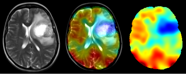
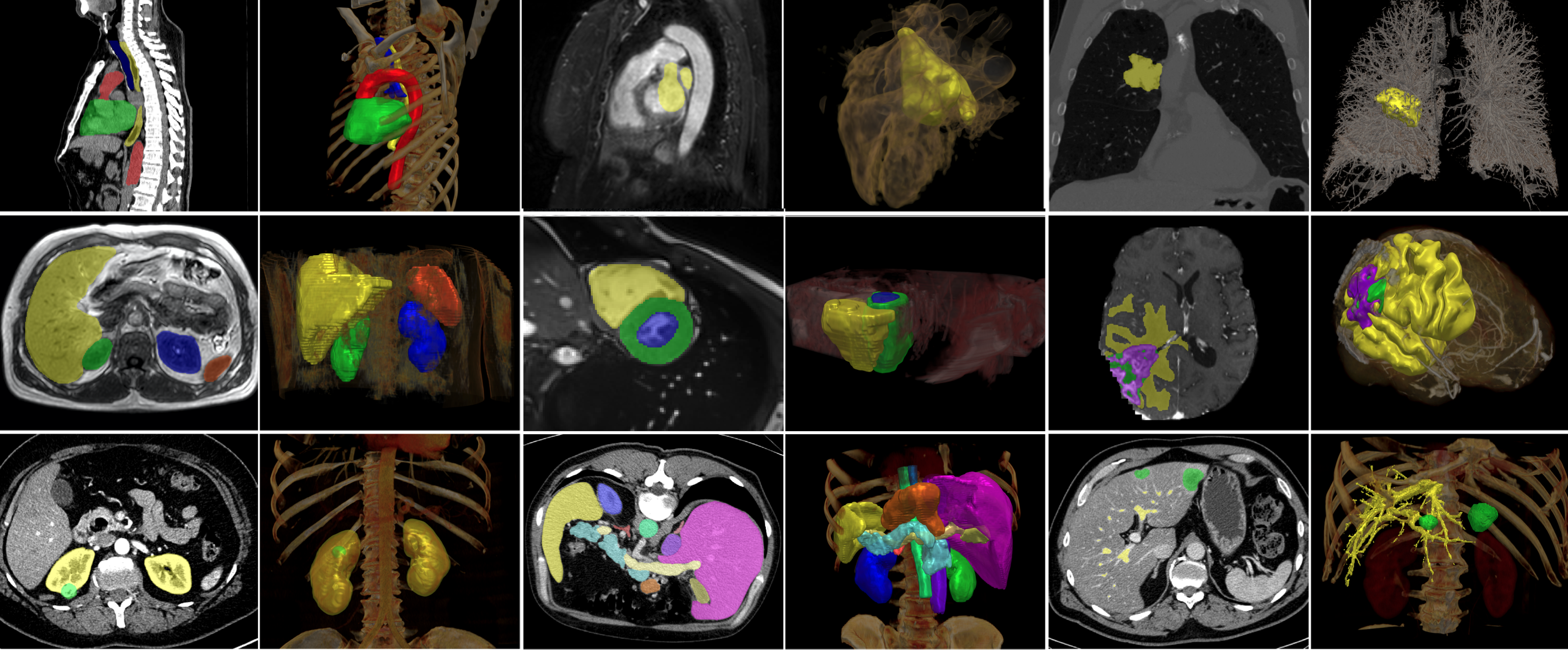
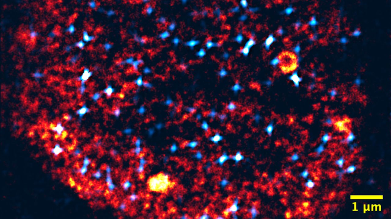
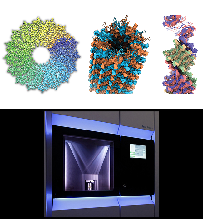
The application areas of imaging in life sciences and soft matter are almost inexhaustible in number. Highly specialized techniques and methods have thus been developed over time for imaging and analyzing a wide variety of samples, giving us unique insights into their structures at all scales.

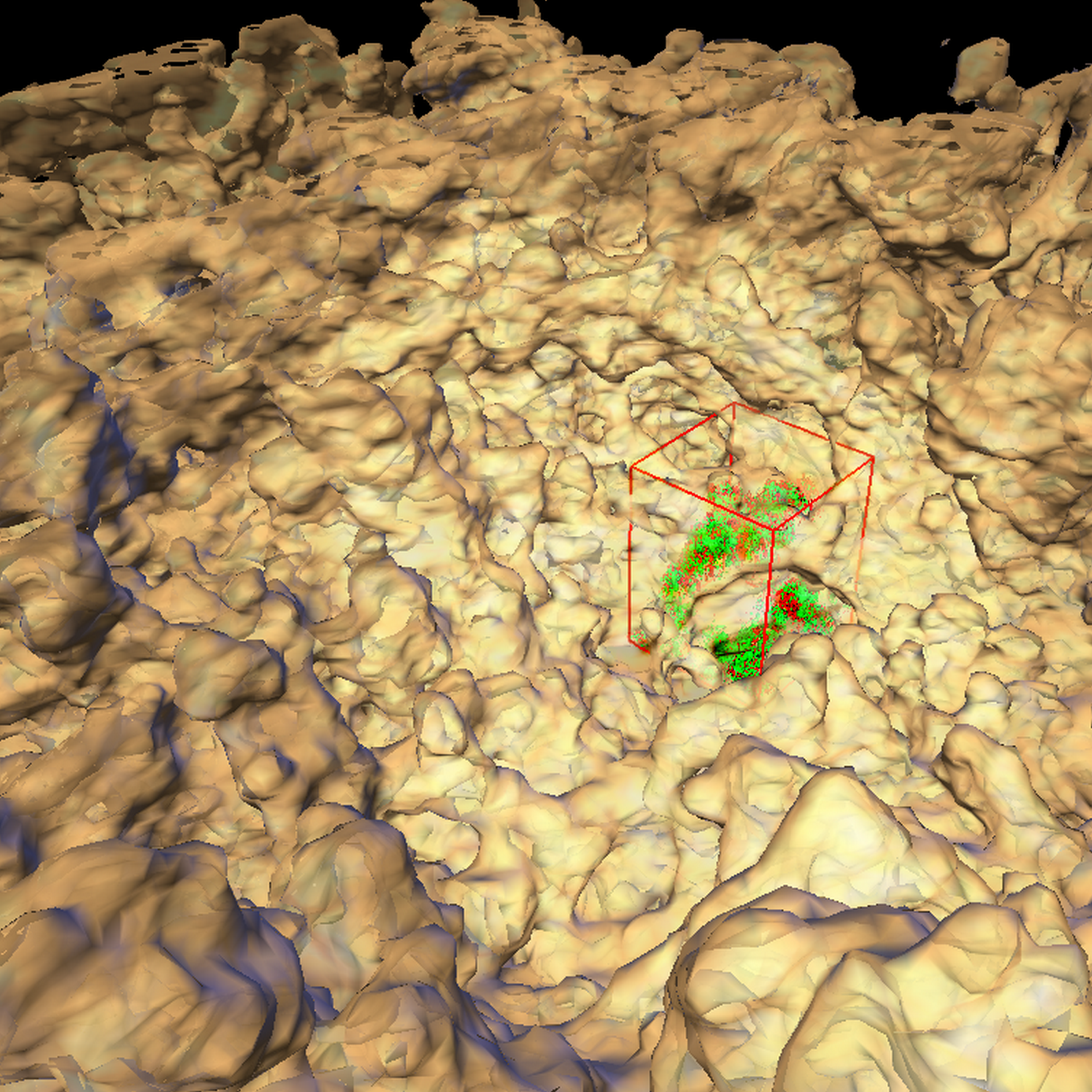
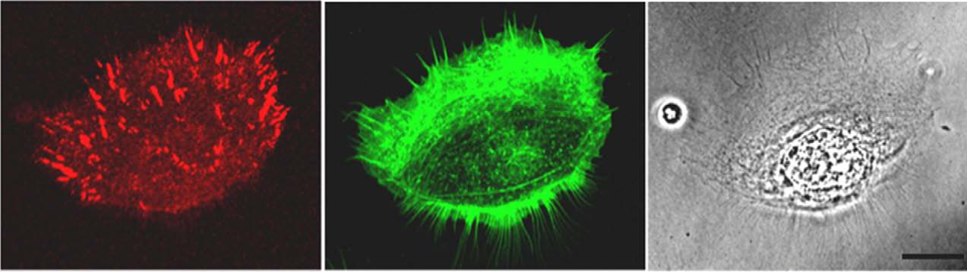
Over the course of this lecture series, data scientists and domain scientists will introduce you to a great number of imaging techniques. Starting with techniques for the study of organisms (X-ray imaging, CT, PET, ...), we address techniques that allow for increasingly fine resolution. Continuing with optical coherence tomography and a variety of microscopy techniques (fluorescence microscopy, LSM, ...) for analysis on the level of cell size, we complete the series of talks with electron microscopy to image structures in the single-digit nanometer range. The speakers will highlight both physical aspects of imaging as well as user-specific issues and topics of data analysis.

This lecture series is held by PIs of the organizing schools and Helmholtz Imaging. The course consists of lecture-style elements and interactive discussions. It covers different imaging techniques that are widely applied in life sciences and soft matter. For an in-depth understanding of the subject, we recommend attending all lectures. Some lectures build on each other, but are self-contained in general. Registration is mandatory for participation. Places are limited and in the case of overbooking, priority will be given to fellows (members) of the three schools. Participants demonstrating and attendance of more than 75% attendance, you can receive a certificate of participation.

Dates: Thursdays, 14-15:30.

Information on the dates, speakers and lecture titles can be found below.

Organization & Registration

The lecture series is offered online. It takes place (more or less) bi-weekly. Registration is required, a registration link can be requested via mail to sabine.niebel@helmholtz-imaging.de

Workshop on Image Analysis

Following the lecture series, there will be a short workshop on image processing to complement the theoretical and methodological content of the lectures. An image viewer software will be presented that allows a lot of plugins which facilitate scientific image analysis. The focus will be on hand-ons and practical exercises.

The exact date, content and registration link for the workshop will be announced in the lecture.

                    Image Lecture Ladd        Image Lecture Nienhaus   


This event is organized by

International Helmholtz Research School of Biophysics and Soft Matter    |    BioInterfaces International Graduate School

Helmholtz Information & Data Science School for Health    |    Helmholtz Imaging

    • 1
      Imaging Physics in Medicine

      Macroscopic imaging in medicine is used both for clinical diagnostics and therapy planning in human patients as well as in research, where both human and small-animal imaging play an important role. Medical imaging is based on several physical principles ranging from ultrasound to ionizing radiation coming from sources outside or inside the patient. Accordingly, the questions to be answered by the different methods and the information in the resulting images are quite different. In this lecture, the physical and technical principles of techniques such as computer tomography (CT), positron emission tomography (PET), and magnetic resonance imaging (MRI) will be introduced.

      Speaker: Prof. Mark Ladd (Medical Physics in Radiology, German Cancer Research Center (DKFZ))
    • 2
      Analyzing Medical Images Using Machine Learning

      The success of Machine Learning has revolutionized the field of medical image analysis in the past 5 years. This talk will give an introduction to relevant concepts in machine learning with a focus on computer vision. Subsequently, several example applications in the biomedical domain will be discussed to study the current state of research and the associated challenges and opportunities.

      Speakers: Lukas Klein (Helmholtz Imaging, DKFZ), Dr Paul Jäger (Helmholtz Imaging, DKFZ)
    • 3
      Optical Coherence Tomography in Biofilm Research - Visualization and Characterization of the Mesoscopic Biofilm Structure

      Understanding the biofilm structure is important in order to understand the structure-function relations of biofilm systems. For decades microscopic tools have widely been applied visualizing the microscopic structure of biofilms (e.g., distribution of cells within the matrix of extracellular polymeric substance). However, mass transport and transfer occurs at a larger scale (mesoscale, mm-scale). By applying optical coherence tomography (OCT) the mesoscopic biofilm structure is assessed non-invasively, in situ and with almost no sample preparation. OCT thus has become popular in biofilm research within the last 10 years.
      This lecture will introduce OCT and provide insights into the technology and possible outputs (e.g., structure analysis).

      Speaker: Dr Michael Wagner (KIT)
    • 4
      Basics of Light Microscopy for the Study of Cells

      Light microscopy is one of the most important techniques for the investigation of animal cells. In this lecture I will present basics of widely used light microscopy techniques including fluorescence and confocal fluorescence microscopy as well as their application on living cells.

      Speaker: Prof. Rudolf Merkel (FZJ)
    • 5
      Advanced Fluorescence Microscopy

      After a brief, general introduction into fluorescence microscopy, I will focus on three modern modalities, STimulated Emission Depletion (STED) nanoscopy, Single-Molecule Localization Microscopy (SMLM) and Light Sheet Microscopy (LSM). STED and SMLM are powerful techniques for cellular imaging with nanoscale spatial resolution, whereas LSM is an excellent and versatile imaging method with diffraction-limited resolution and low phototoxicity. Pros and cons of the methods for particular imaging applications will be discussed.

      Speaker: Prof. Gerd Ulrich Nienhaus (Institute of Applied Physics, KIT)
    • 6
      Microscopy Assessment of DNA-based Information Processing in Biological and Artificial Systems

      The processing of DNA sequence information is a central task of biological cells, and could in the future enable molecular biotechnologies with “smart capabilities”. In our research group, we combine cutting-edge light microscopy, computational data analysis, and simulations to understand the principles of this information processing in the 3D-space of the cell nucleus. In this lecture, I will outline how such imaging-informed assessments can reveal major principles of DNA-based information processing, and how these principles might in the future guide the design of artificial, DNA-based information systems.

      Speaker: Prof. Lennart Hilbert (KIT)
    • 7
      Imaging Biological Molecules by Electron Cryo-Microscopy (cryo-EM)

      The cryo-EM method has become very powerful in the past several years and it is becoming the method of choice for many structural biologists. The interpretation of single noisy molecular images in solution still presents an obstacle for obtaining faithful structural information of biological macromolecules. Therefore, we routinely apply image processing routines of structural averaging in single-particle cryo-EM as well as cryo-tomography. Small quantities of purified proteins and protein complexes can be visualized in vitreous ice using single-particle cryo-EM. Subsequent data processing of 1000s of molecular images enables three-dimensional structure determination at near-atomic resolution. The obtained atomic models provide the chemical framework for biological macromolecules with respect to their biological function. Alternatively, more complex biological environments such as cells are increasingly being visualized by cryo-EM methods. Electron cryo-tomography combined with fluorescence light microscopy enables the detailed characterization of natively preserved cells, the organelle ultrastructures and provides the link to their molecular interpretation. In the talk, we will explain the physical foundations of the cryo-EM techniques and associated image processing approaches and combine them with recently developed applications that assist in the interpretation of the native electron cryo-micrographs.

      Speaker: Prof. Carsten Sachse (Forschungszentrum Jülich GmbH, Ernst-Ruska Centre for Microscopy and Spectroscopy with Electrons / ER-C-3 Structural Biology, Institute for Biological Information Processing / IBI-6 Cellular Structural Biology)