Choose timezone
Your profile timezone:
ATTENTION! On Wednesday, October 22 at 10:00 a.m., we will delete all events older than 6 months from the Test category. If you have regular events there, please move them in time to prevent data loss!
For some registered participants, mails from this platform seem to end up as spam. To make sure to receive all information, please check your junk folder.
Per default all times are given in CEST. To make sure to refer to your local time, please set your time zone accordingly. Click the menu at the top right of this page.
After a series of successful BigBrain workshops we are pleased to announce the 5th BigBrain Workshop, taking place as an international symposium on:
September 22-23, 2021 in virtual format
With a pre-workshop day on September 21 providing practical hands-on sessions on our data and tools.
We are proud to announce Martijn van den Heuvel as our keynote speaker, talking about 'The brain network - from cell to macroscale circuits to function'.
The full program will be released here soon. Stay tuned.
While it was originally planned to take place as a physical event in Zadar, Croatia, we were unfortunately compelled to move to an entirely virtual event again this year. We hope to be able to make up for this next year.
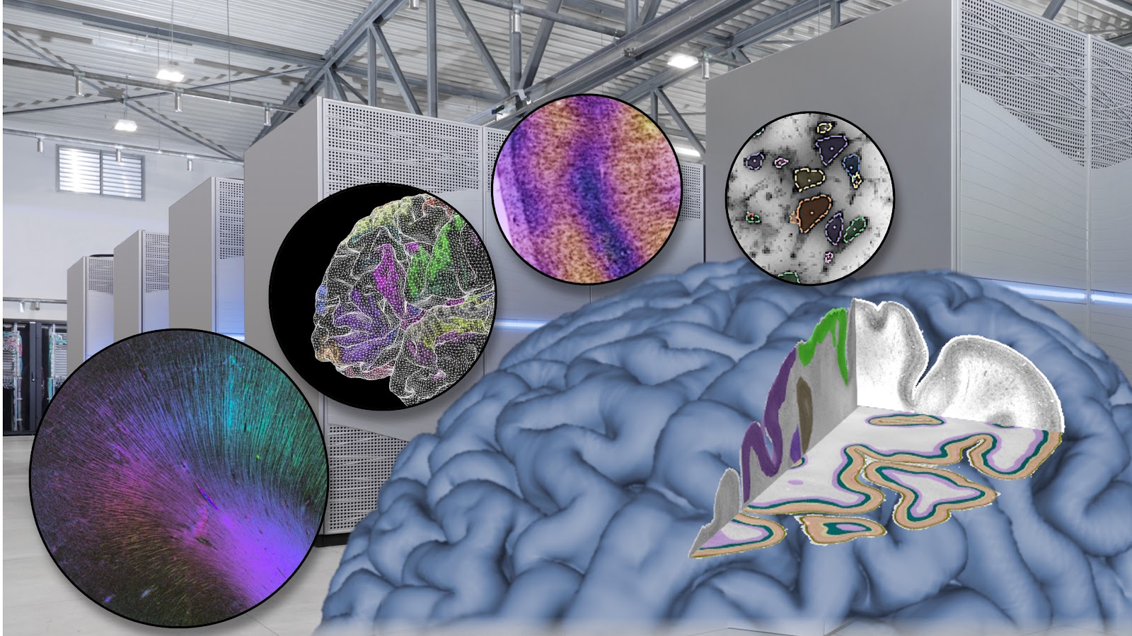
We are delighted that the cooperation between McGill University in Montréal and Forschungszentrum Jülich, one of our longest and most successful partnerships, has now been formally organized as Helmholtz International Lab. This enables us to increase our efforts to build the next generation of multimodal human brain atlases around the successful BigBrain model. With HIBALL we develop highly detailed 3D brain models using novel AI methods and supercomputing architectures. These models will be exposed through a sustainable, transcontinental research platform for computation and data sharing, with high interoperability with the systems of large brain initiatives. With the upcoming BigBrain Workshop we aim to reach out to the international community of BigBrain users and to invite researchers from our global network (and beyond) to present their work and to discuss future prospects of the BigBrain data and tools.
The event is free of charge but prior registration is required. Please register by September 20, 2021 at the latest.
The workshop will be organized as a symposium, with both invited speakers and contributed talks as well as a poster and demo session. We welcome short abstracts of current work and/or short proposals for future initiatives related to the BigBrain. The topics to be considered will include:
All talks will be recorded and published on bigbrainproject.org.
Please contact the program committee if you have any questions. We will continuously update the information on this page and also share information via Twitter (@BigBrainProject) and e-mail.
![]()
![]()
![]()
![]()
The goal of this course is to familiarize scientists with no, or only very little, knowledge about brain anatomy with major brain structures and their functions. You will learn about the landmarks used by anatomists to navigate through the brain, and the functions they are involved in. Most importantly, you will have a chance to search for these structures yourselves. By the end of the course you will not only be familiar with terms such as lobes, gyri, diencephalon, hippocampus or visual cortex, but you will also be able to find these structu
High resolution histological data provides a unique perspective on the cellular structure of the brain. Histological data is available for a large number of species, and the possibility of staining for particular aspects of the tissue allows the researcher to formulate an extremely rich range of questions. It presents, however, several challenges which make its analysis difficult. In particular, the data is affected by various types of artefact, and the subtle differences that distinguish one structure from the other require a well trained human eye. In addition, scanned at very high resolution, the file sizes involved become difficult to manipulate. These may be in part the reasons why the expert segmentation of histological material is often performed by a single researcher.
MicroDraw is an online tool for the collaborative segmentation of high-resolution histological data. MicroDraw uses deepzoom to enable rapid access to high-resolution data without limits in image size. Images can be manipulated in any Web browser, in computers, tablets or even smart phones. MicroDraw provides a growing number of tools for vectorial annotation, which allow us to segment data at any resolution. MicroDraw greatly simplifies distributed collaboration, by providing researchers access to the same dataset independently of the computer where the data is hosted. MicroDraw provides simple tools for the definition of collaborative projects, and helps coordinate access to data and results. Finally, MicroDraw implements a RESTful API, which allows researchers to programmatically query the segmentations performed in a project and use sophisticated image analysis tools for their analysis.
In this tutorial we will show how to encode data and host it to make it accessible. We will use the BigBrain data as an example. We will then show how to visualise data and annotate it using the different vectorial annotation tools. We will show how to create a project to centralise a set of annotations. Finally we will show how to query that data using a Python script, display the data obtained and compute some simple measurements.
MicroDraw is open source and we invite you to contribute to its development either as a user or as a developer.
This tutorial will provide an introduction to two tools that can be used to process and manage BigBrain-related data: CBRAIN and DataLad.
CBRAIN is a web portal that provides seamless access to high-performance computing clusters. DataLad is a data integration tool to keep track of distributed datasets. The tutorial will cover the main functionalities of CBRAIN and DataLad, illustrate them on BigBrain data, and demonstrate their interaction.
The EBRAINS multilevel human brain atlas integrates maps that capture different facets of human brain organization into a common framework. It is defined across multiple reference spaces, where BigBrain represents the micrometer level. The atlas links cytoarchitectonic areas with ultra-high resolution BigBrain data, complements them with maps of fibre architecture and functional organization, and links them to a growing set of multimodal data features associated to brain regions. EBRAINS atlas services are designed as an online framework that can be accessed interactively using an online viewer, as well as in a programmatic fashion through structured Python and http interfaces. This tutorial will give a tour of the recent status of EBRAINS atlas services, and discuss some future directions.
The BigBrain Warp is a toolbox for multi-modal integration of BigBrain, composed of a centralised repository of BigBrain related transformations and scripts to easily move between histological and MRI spaces. In this session, we'll walkthrough the toolbox and guide short tutorials on how to use BigBrain in the context of structural and functional MRI.
Cytoarchitecture is defined as the spatial organization of neuronal cells in the brain, including the arrangement of cells into layers and columns with respect to cell density, orientation and presence of certain cell types. It allows to subdivide the brain into cortical areas and subcortical nuclei, which are indicators for connectivity and function. Consequently, cytoarchitectonic areas provide an important microstructural reference for human brain atlases.
Today's high-throughput scanners enable digitization of complete human brains in reasonable timeframes, opening up oppurtunities for large scale analysis of cytoarchitecture. However, it is practically impossible to scale established cytoarchitectonic mapping methods for doing delinations in all sections of a human brain. This motivates the development of automatic mapping algorithms.
In this talk, we discuss the challenges of automatic cytoarchitectonic mapping, give an overview of recent advances in the field, and talk about potential future developments.
The visual system of mammals is comprised of parallel, hierarchical specialized pathways. Different pathways are specialized in so far as they use representations that are more suitable for supporting specific downstream behaviours. In particular, the clearest example is the specialization of the ventral (“what”) and dorsal (“where”) pathways of the visual cortex. These two pathways support behaviours related to visual recognition and movement, respectively. To-date, deep neural networks have mostly been used as models of the ventral, recognition pathway. However, it is unknown whether both pathways can be modelled with a single deep ANN. Here, we ask whether a single model with a single loss function can capture the properties of both the ventral and the dorsal pathways. We explore this question using data from mice, who like other mammals, have specialized pathways that appear to support recognition and movement behaviours. We show that when we train a deep neural network architecture with two parallel pathways using a self-supervised predictive loss function, we can outperform other models in fitting mouse visual cortex. Moreover, we can model both the dorsal and ventral pathways. These results demonstrate that a self-supervised predictive learning approach applied to parallel pathway architectures can account for some of the functional specialization seen in mammalian visual systems.
For the detection of neuronal cell bodies in 1-micron BigBrain data we propose a conceptually simple framework called Contour Proposal Network (CPN). The CPN detects and segments possibly overlapping cells by fitting closed contours using a fixed-sized representation based on Fourier Descriptors. State-of-the-art object detection architectures can be used as backbone networks, forming a single-stage instance segmentation model that is trained end-to-end. We evaluate the CPN with different backbone networks using datasets from different modalities, including the 1-micron BigBrain. Experiments show that CPNs outperform U-Net and Mask R-CNN in instance segmentation accuracy. The CPN is computationally very efficient and is suitable for real-time applications when coupled with backbones such as ResNet-50 FPN. The trained models generalize well, even across different domains of cell types. The main assumption of the method regards closed object contours, hence the CPN is applicable to a wide range of detection problems also outside the biomedical domain. PyTorch code has been made available at: celldetection.org
Low-rank-based representation learning is powerful for recovering the subspace structures in data, which has obtained an impressive performance; however, it still cannot obtain deeply hidden information due to the essence of single-layer structures. Structure and nonlocal patch similarity have been used successfully to enhance the performance of image restoration. However, these techniques can often remove textures and edges or introduce artifacts. In this article, we investigate the deep low-rank representation of images by presenting a novel strategy that can extend existing single-layer latent low-rank models into multiple layers. Technically, we propose the Deep Latent Low-rank learning and global structure sparsity for Bigbrain super-restoration to uncover deep features. Extensive results on the databases show that our framework can deliver enhanced performance over other related techniques.
The medial geniculate nucleus (MGB) is part of the metathalamus and plays an important role in processing auditory information. Previous maps of the human brain did not include subdivisions of the MGB – limiting their use for data integration, modelling and simulation. Here we aim at overcoming this limitation by creating cytoarchitectonic maps of the MGB in the BigBrain (Amunts et al., 2013) with the help of a deep-learning based brain mapping tool (Schiffer et al., 2020). In a first step the MGB was analyzed on 57 sections of the BigBrain dataset based on cytoarchitectonic criteria. Three subdivisions were identified and mapped in both hemispheres. In a next step, a deep-learning based tool with a convolutional neural network architecture was trained to delineate these subdivisions on additional 132 sections of the BigBrain. After an initial quality check, the maps were non-linearly transformed into the 3D BigBrain space and smoothed. The resulting whole brain maps serve as a histological reference for clinical and neuroscientific research investigating medial geniculate function and accompany recent advances in spatial resolution of in vivo imaging techniques therein. The maps are publicly available and can be accessed via the EBRAINS Knowledge Graph and the Human Brain Project’s interactive atlas viewer (https://interactive-viewer.apps.hbp.eu/).
The nucleus ventralis intermedius (VIM) of the thalamus is located in the ventral part of the ventrolateral posterior thalamus and exhibits increased connectivity especially with the dentate nucleus of the cerebellum and the primary motor cortex through the dentato-rubro-thalamic tract. Selective lesioning of the VIM is a well-established neurosurgical target for the treatment of patients with severe medication-resistant tremor as it leads to a significant alleviation of this symptom. Most current image-guided techniques are based on indirect targeting using atlases (e.g. Schaltenbrand-Wahren atlas) for determining and navigating the strategy of the surgery, which has certain limits of spatial resolution and contrast. Since the precise targeting of VIM and a comprehensive understanding of its inner structure is important for the success of neurosurgical treatment we investigated the VIM in serial histological sections of ten human post mortem brains and mapped the structure according to cytoarchitectonic criteria at every 15th section. The BigBrain2 dataset was one of the investigated brains and was used to train a deep-learning model that learned to recognize cytoarchitectonic patterns based on manual delineations and predicted the delineations at every section in-between. VIM was then visualized to present a detailed anatomical model of the human VIM at microscopical resolution in order to better understand the 3D architecture and to get an overall picture of its shape and relationship to other nuclei of the thalamus.
The current state of reconstruction of BigBrain2 is presented, describing
the brain of a 30 year old anonymous male donor. As for the original BigBrain,
cell-body stained histological coronal sections were digitized and
reconstructed in 3D. Sections are first repaired at 20 microns in-plane
resolution to correct for manipulation artifacts, then aligned to the MRI
serving as the undistorted frame of reference. Given the time-consuming
nature of the manual aspects of the corrections, including provenance
tracking of the operations for reproducibility and variability assessment
in section repairs, every fifth section was initially repaired and a first
optically-balanced aligned volume at 100 microns isoptropic resolution was
obtained. This preliminary 3D alignment at 100 microns defines a transformation
to the MRI which can subsequently be used for the newly repaired sections being
added to the pipeline (25% completed). The transformations defined at 100 microns
will be applied at full resolution to obtain a 3D volume at 20 microns
isotropic resolution prior to performing non-linear section-to-section
alignment at finer resolution levels once all sections have been repaired.
This multi-resolution approach can be extended for future reconstructions
of BigBrain3 at full 1 micron resolution by first downsampling the 1 micron
sections to 20 microns. With provenance tracking, manual repair operations
carried at 20 microns can be reproduced at the 1 micron level.
Mapping the microscopical organization of the human brain provides an important basis for multimodal brain atlases, and is indispensable for linking functional, physiological, connectivity, molecular, or genetic properties to their cellular correlates. The BigBrain (Amunts et al., 2013) is a 3D model of a complete human brain at microscopic resolution, constructed from more than 7000 histological sections at a resolution of 20 micron isotropic. By resolving cortical layers, subcortical nuclei, and even larger individual cell bodies, it has enabled a new generation of high-resolution studies. Yet, the resolution of 20 micron is not sufficient to perform classical cytoarchitectonic mapping in arbitrary cutting planes, or quantitative analysis of 3D distributions and numbers of individual cell bodies. For such types of analyses, the construction of 3D brain model at the resolution of 1 micrometer is mandatory. While recent technologies in high-throughput microscopic imaging, large-scale storage, and high-performance computing have brought such an endeavour into sight, several challenges need to be addressed to compute such a model. These range from distributed data management to new image registration paradigms, very large numerical optimization problems, to cloud technologies for providing remote access to image data in the Petabyte range. In this talk, we will describe some of these challenges and recent progress on feasible solutions.
We present a 3D reconstruction pipeline for 2D autoradiographs that will allow for the creation of the first ever set of ultra-high resolution (50𝜇m). The 3D atlases for 20 different neurotransmitter binding sites in the human brain. This pipeline was designed to overcome significant challenges in the data, including: non-linear deformations in the brain tissue, intensity variations between autoradiographs, variability in autoradiograph acquisitions, a large number of slices lost in sectioning, and non-orthogonal brain sections. Moreover, because sections are serially sectioned for 20 different ligands, there are significant gaps between autoradiographs for a given ligand. Surface-based linear interpolation was used to interpolate ligand densities over the cortex. Surface ligand binding densities were interpolated into a 3D volume to create atlases of ligand binding densities.
Finally, the two methods have been implemented to validate the quantitative accuracy of the reconstructed ligand values. The first approach involves directly comparing the pixel intensities of sections in the reconstructed ligand volumes to the raw autoradiographs. This is done to verify that the pipeline preserves the ligand binding densities. Initial results indicate ~90% accuracy for a flumazenil binding density volume at 0.6mm resolution.
The second validation technique applies the surface-based interpolation algorithm within acquired autoradiographs. This involves identifying a random set of vertices within aligned autoradiographs and attempting to interpolate the binding densities using the surface-based interpolation algorithm.
The methods presented here provide all the necessary processing steps to reconstruct a dataset of multiple neurotransmitter receptor atlases at 50𝜇m for a full human brain.
Given the usefulness of the BigBrain high resolution histological volume, it would be very valuable to have a similar public domain resource for the macaque. Isotropic high resolution data to build such a resource is not currently available, but as an initial step we present a histological volume based on the NIH Blueprint Non-Human Primate (NHP) Atlas [1], acquired by the Allen Institute and funded by the NIH. Our contribution is to take the set of Nissl stained slices (50 $\mu m$ thick, 250 $\mu m$ apart) and to create a smooth volume by calculating the optimal alignment. We used open tools [2, 3] for processing, modernized the code of the poSSum three-dimensional reconstruction toolbox [4] and extended the deformable registration method thereof, using the an MRI-based macaque atlas [5] as a reference template. We will use the reconstructed volume to estimate neuron densities across all cortical areas used in the core-nets.org macaque connectivity database [6]. Besides adding to the body of knowledge on the cytoarchitecture [7] and geometry of the macaque cortex, this work will provide tools to further analyze histological data and support large-scale dynamical modeling studies.
[1] NIH Blueprint NHP Atlas, www.blueprintnhpatlas.org
[2] Bakker et al. Neuroinformatics, 2015
[3] Microdraw, microdraw.pasteur.fr
[4] Majka et al. Journal of Comparative Neurology, 2016
[5] Calabrese et al. NeuroImage, 2015
[6] Markov et al. Cerebral Cortex, 2012
[7] Beul and Hilgetag. NeuroImage, 2019
Ex-vivo high-resolution MRI of brain tissue can provide morphological and microstructural information. MR images can also serve as undistorted references for the reconstruction of digitized histology, immunohistochemistry, or clearing techniques. Small-bore scanners, equipped with powerful gradients systems and operating at ultra-high magnetic fields present the optimal conditions for ex-vivo MRI. However, space constraints make it impossible to scan a whole human brain in such small-bore scanners. Imaging a human brain requires a large bore scanner, typically showing limited gradient system performance, constraining the achievable spatial resolution. Importantly, large histology sections cut from large brains are prone to distortions, tears, and folding.
Here we present a method for MRI-guided hierarchical sectioning and stitching of brain blocks for precise alignment of processed tissue images to corresponding MRI volumes. Our method allows the use of small-bore MRI scanners, achieving high-resolution-MRI volumes. The use of small tissue blocks reduces tissue distortions, tears, and foldings. The results show significant improvements relative to current methods for 3D reconstruction of 2D processed tissue images and for aligning processed tissue data to the corresponding MRI volumes.
How does our brain give rise to brain function, cognition and behaviour? Understanding the fundamental features of brain networks (‘the connectome’) and linking the different levels and scales of brain connectivity to each other is key in tackling this challenge. We will start by discussing the organization of brain networks and network theories of how 'principles of wiring’ may shape integration, functional specialisation and diversity. We will particularly take a look at how variation in these connectome properties may give rise to variation in behavior, but also how they may shape a landscape of brain disconnectivity within and across disease. One key factor in understanding brain connectivity (and disconnectivity) is to understand how connectivity is organised across scales, from genes to cells to macroscale circuits to behaviour. Celebrating game-changing initiatives such as the BigBrain project, the Allen Human Brain Atlas and the UKB (among many others) who provide the field with exciting new ways to link connectivity across scales, we will discuss the opportunities that ‘multiscale neuroscience’ can bring to understand brain network organization in health and disease.
The Sievers Computational Neuroscience Initiative (SCNI) builds cross-disciplinary experience in neurological sciences research and neuroinformatics to establish a computational neuroscience approach to modelling brain states in healthy ageing and disease.
The SCNI provides training and mentoring for tomorrow’s researchers to undertake and partake in complex studies, and to build the computer infrastructures and brain-based datasets to support this research.
Martijn van den Heuvel is a multidisciplinary scientist with a background in Cognitive Artificial Intelligence (MSc, 2004) and psychiatric medical imaging (PhD, 2009). His research focus is the network of connectivity of the human and animal brain, the connectome, with the aim to get better understanding of the fundamental rules of wiring of nervous systems, and in particular how these rules of wiring are associated with brain function and disfunction in health and disease. His field of expertise includes structural and functional MR imaging combined with network science, bridging the field of mathematics, informatics, psychology and medicine. He heads the dutchconnectomelab.org at the Brain Center Rudolf Magnus, Utrecht, The Netherlands. Martijn received Dutch Research Council NWO-VIDI and NWO-VENI awards, an international MQ Fellowship (www.joingmq.org), and the 2013 Dutch Brain Trophy of the Dutch Brain Foundation.
Background. Complex behaviours benefit from parallel distributed processing in multiple brain networks. The roles of certain networks are well-defined, while others remain elusive. Arguably, none are so elusive as the default mode network (DMN); a distributed set of brain regions that decrease in activity during many externally oriented tasks. Revealing the cytoarchitectural composition and connectional layout of the DMN is crucial to defining its role in complex behaviours.
Method. We examined the cytoarchitectural composition of the DMN using an established cortical type atlas (García-Cabezas et al., 2020; Von Economo and Koskinas, 1925) and by applying non-linear dimensionality reduction to BigBrain-derived staining intensity profiles (Paquola et al., 2019). Next, we used magnetic resonance imaging (MRI) to explicate structural wiring and effective connectivity of the whole brain. In both modalities, we examined the influence of cytoarchitecture on extrinsic connectivity of the DMN. Finally, we evaluated the uniqueness of the DMN relative to other large-scale functional brain networks.
Results. We discovered profound diversity of DMN cytoarchitecture. Each circumscribed subregion of the DMN contains a broad range of cytoarchitectural types, however, the spatial pattern within each subregion differs. The patterns vary in smoothness from a gradient in the parahippocampus to interdigitation in the superior frontal gyrus. We found that cytoarchitectural differentiation in the DMN aligns with its structural wiring and extrinsic information flow. The structural heterogeneity of the DMN engenders a network-level balance in communication with external and internal sources, which is distinctive, relative to other functional networks.
Conclusion. These findings suggest a novel wiring diagram of structural and functional connectivity of the DMN that is compatible with its putative role in balancing internal and external information. Furthermore, our work demonstrates the import of neuroanatomical evidence in specifying theories of functional networks.
Analysis of histological data requires sophisticated methods and tools. In the past, research teams have aimed at producing tool packages covering all researchers’ needs. Such large packages can quickly become unwieldy: difficult to maintain and complex to use. Web technologies allow for a powerful alternative: focused micro-services. Instead of a single, overly complex tool, users can rely on a variety of small tools, easier to develop, easier to understand, each aiming at solving a single problem in an efficient manner. For the same problem, different users may have the possibility of picking the tool that is the most appropriate to their working style.
We demonstrate the possibility of creating such services using MicroDraw, a Web app for the collaborative annotation of high resolution histological datasets (https://microdraw.pasteur.fr). We developed a website implementing an external service for segmenting histological data using a random forest classifier. A series of sample regions can be delineated in MicroDraw , and fetched by our service. The website uses Pyodide, allowing us to run Python machine learning code in the web browser. We used Scikit-image for extracting image features, and Scikit-learn for the classification algorithm. The regions used to train the classifier are adjusted iteratively to optimise the segmentation, and applied automatically to a series of images of the same type. The results are finally vectorised and sent back to MicroDraw. More generally, the same procedure could be used with different machine learning or deep learning algorithms, facilitating the interconnection of different services, fostering distributed collaboration.
Connections and interactions among neurons manifest as patterned neural activity and adaptive behaviour. Ascending projections from the brainstem and subcortical nuclei have a modulatory effect on the electrical potential - and therefore the excitability and firing rate - of cortical neurons (Shine 2019). These modulatory influences are coordinated by overlapping and heterogeneous distributions of multiple neurotransmitter receptors at the target cells. This heterogeneous distribution of neurotransmitter receptor densities across the cortex suggests a diversity of modulatory influence, and therefore also of signal integration, neural dynamics, and whole-brain connectivity. We used two recent state-of-the-art datasets (PET and autoradiography) of neurotransmitter receptor densities across the neocortex, which include a total of 32 excitatory, inhibitory, ionotropic, and metabotropic neurotransmitter receptors, transporters, and receptor binding sites (Zilles et al., 2017). First, we mapped receptor distributions to structural and functional connectivity, thereby profiling how receptors may influence whole-brain communication. Second, we used multiple linear regression models to predict MEG-derived neural dynamics from receptor densities and find that excitatory ionotropic receptors are dominant contributors toward shaping neural dynamics. Finally, we asked how neurotransmitter receptor densities map onto meta-analytic patterns of functional activation from Neurosynth (Yarkoni et al., 2011) and disease-specific cortical thinning from the ENIGMA consortium (Thompson et al., 2020). Altogether, we uncover the neurochemical infrastructure that shapes the brain's connectivity and dynamics by comprehensively mapping receptor distributions to the structure and function of the human brain.
Extensive research over the past three decades have been revolutionary by conceptualizing a neuron-glial paradigm which aims at describing the mutual dependence between glial and neuronal processes at multiple spatiotemporal scales. However, glial contributions to large-scale functional neuronal network organization remain mysterious due to the lack of empirical and theoretical frameworks elaborating on neuron-glial interaction mechanisms at a macroscopic scale.
Here, we developed a biophysical neuron-glia mass network modeling approach to explore how variations in glial network activity could reshape emergent brain-wide neuronal functional connectivity patterns.
Our model explains local dynamics by coupling bilaterally neuronal and glial activity though the modulation of extracellular glutamate and GABA concentrations. In addition, our model assumes that glial masses interconnect only to their first neighbors along the cortical mantle, while neuronal masses interconnect through white matter tracts as empirically derived from diffusion magnetic resonance imaging tractography.
By tuning model parameters that control the relative contributions of glial network activity in inducing glutamate and GABA neuronal release by acting on presynaptic neurons, we simulated multiple whole brain activity with distinct spatiotemporal signatures. We used phase-locking value to quantify the synchrony patterns between neuronal populations thereby providing neuronal functional connectomes, and we used graph theoretical indices to describe the topological properties of these connectomes.
We report a non-trivial dependence between glial network induced changes in excitatory and inhibitory synaptic transmission and neuronal functional connectivity. Importantly, we provide a new perspective of functional organization and operation of neural networks, inclusive of glial processes.
Segmentation of the human cerebellar cortex from histological data has been considered a challenge due to its convoluted structure, and the numerous artifacts caused by sectioning or staining. The high resolution BigBrain model (Amunts et al., 2013) enables to overcome the lack of detail occasioned by the limited number of sections in existing datasets, and the large BigBrain data is ideal for developing AI solutions for segmentation. One would normally need a large number of annotated labels for deep learning-based models such as Convolutional Neural Networks (CNNs) to perform accurate segmentation. However, anatomical annotation needs expert-level knowledge, and a sheer volume requirement on the annotation would hinder the application for cerebellum segmentation.
In this work, we seek to leverage domain adaptation on existing annotated cerebellum data from the Allen Brain atlas (https://atlas.brain-map.org) to ease the demand for annotations of BigBrain. Here we demonstrate how we pre-train a segmentation model for the cerebellum on the existing Allen Brain labels and generate pseudo labels for our BigBrain data to train a new segmentation model. The Dice loss between the predicted segmentation results and pseudo annotations will serve to pick 50 data with the highest loss to conduct manual annotations, which will then be used to train the final segmentation model.
Our proposed framework aims to combine active learning with domain adaptation for an annotation-efficient cerebellum segmentation.
Reference brain cortical surfaces derived from various structural pipelines enable integration of multimodal data into a standard space. In the absence of a common framework across structural pipelines, high profile surface atlases created within FreeSurfer (fsaverage) or Human Connectome Project (fs_LR) are not available in standard reference frames like the MNI152 or the 3D-reconstructed histological BigBrain model (Amunts et al. 2013). Here, we present our improved surface registration pipeline linking the BigBrain surface with other reference surfaces of interest (Lewis et al. 2020).
We implement a reparameterized multiscale pipeline via the Human Connectome Project's (HCP) Multimodal Surface Matching (MSM) tool (Robinson et al. 2014, 2018) and HCP workbench (Marcus et al. 2011). The BigBrain surface (Wagstyl et al. 2020) is first re-tessellated using mris_remesh (FreeSurfer7.1), which eliminates the suboptimal unfolding of the right occipital pole observed with our previous version. Registration is then carried out in a direct manner from the re-tessellated BigBrain surface to the reference surface. Performance of the updated pipeline shows improved accuracy and comparably low distortion as our previous approach.
This work allows the high-resolution, histological BigBrain model to serve as an unprecedented cross-validation tool for surface registration pipelines. Any surface atlas defined in another standard space, e.g. fs_LR or fsaverage, can now be transposed to BigBrain space such that macroscopic parcellation boundaries derived from in vivo imaging can be directly compared to cytoarchitectural properties. Likewise, BigBrain's histological landmarks or cortical layers can be transposed to fs_LR and fsaverage for a wide range of functional applications.
Magnetic resonance imaging (MRI) studies show that the majority of the T1-weighted contrast stems from myeloarchitecture, but cytoarchitecture still has an impact on the signal. Many cortical MRI markers are thought of as being driven mostly by myelin. Here, we compare the spatial organization and age effects of cortical thickness (CT) to 3 measures of cortical microstructure (gray-white matter contrast [GWC], the boundary sharpness coefficient [BSC], gray matter [GM] T1w/T2w ratio, and superficial white matter [SWM] T1w/T2w ratio), and probe their specificity to cellular organization using the histological reconstruction of BigBrain.
For each marker and vertex, the mean value across the 127 healthy subjects (aged 18-81, 76F, 51M) was computed and a linear model with age and sex as predictors was fitted. The markers were also generated on the BigBrain volume. The correlation of the surface maps was assessed and significance tested via spin tests.
Overall, results showed higher correlations between spatial distributions than between age trajectories. The GWC and BSC showed significant positive spatial correspondence with their homologous measures derived using BigBrain, while T1w/T2w ratio measures did not.
Our finding of spatial correlations being higher for mean values than for age effect indicates that some measures tend to covary at the cortex-wide level, but age trajectories are likely influenced by different interactions of microstructural changes. Our BigBrain results indicate a general trend of GM T1w signal and myelin being inversely related to the density of cells, although that relationship is weak to moderate.
We propose a 3D surface extraction benchmarking tool to evaluate the performance of 3D model extraction methods from 2D discrete label maps. We have used one geometrical and one anatomical model as references. The anatomical model is selected from the BigBrain hypothalamus atlas (Jones et al. in prep).
3D triangular meshes are used as ground truth 3D surfaces. These contain various geometric features, such as concave and convex curvatures, textures, smooth surfaces, etc. 2D label maps of these reference models are extracted using Atelier3D software (A3D; Borgeat et al., 2007) and 3D surfaces are reconstructed from these using a surface extraction algorithm applying two methods: method 1 uses 2D label maps of every 20 sections, and method 2 includes every 2 sections from the BigBrain sectioning planes to reconstruct the shape of the structure. The reconstructed model is then evaluated by comparing to the original (reference) 3D model, considering features such as Hausdorff distance, RMS (root mean square), volume, surface area, number of vertices, and number of polygons.
Increasing the resolution (by including more sections) improves the performance of 3D surface extraction. This tool allows for interoperability of common visualization and annotation tools used around BigBrain, and provides the ability to compare the performance of different algorithms for 3D model, surface, or mesh creation processes from 3D volumes/2D discrete label maps at different resolutions. Such a benchmarking approach facilitates collaboration, helps improve the accuracy and scalability of 3D surface extraction, and promotes reproducible research.
The expansion of the cerebro-cerebellar system may be a primary driving factor behind primate cognitive evolution. Due to extensive connectivity to cognitive parts of the neocortex, the cerebellum is thought to support cognition analogous to its role in motor functions. Using an extensive MRI dataset (34 primates; 65 specimens) we found that cerebellar and neocortical measurements argue for Brownian Motion evolutionary dynamics. Furthermore, using phylogenetic generalized least squares analysis, we found that cerebellar and cerebral volumes co-evolve in primate evolution. We discuss these findings in reference to comparative neuroscience literature, providing a broader perspective of progress in this field and the usefulness of open science tools to aggregate anatomical data and segmentation.
The cerebral cortex consists of distinct layers with unique properties and functions. Previous work has shown that the laminar architecture of cortical regions varies in a spatially ordered fashion along a ‘‘sensory-fugal’’ axis, with decreasing laminar differentiation from the unimodal to transmodal areas. Indeed, the “Structural Model” proposes that the isocortex can be divided into regions with comparable laminar structures, or cortical types, with links to their connections, plasticity and development. In this study, we leveraged the BigBrain map of cortical layers and used a non-linear manifold learning approach to probe along which organisational axes laminar structure covaries in the cortex
Brain structure scaffolds intrinsic function, supporting cognition and ultimately behavioral flexibility. To evaluate the association between structure and function we assessed whether regions with similar microstructure would also be functionally connected in humans and macaques. This microstructural profile similarity approach has been previously developed in the big brain (Paquola, 2018). In humans, structure-function coupling was highest in regions of unimodal cortex and lowest in transmodal cortex. Structure-function uncoupling in non-human primates had a similar spatial distribution, but we observed an increased coupling between structure and function in association regions in macaques relative to humans.
In current neuroimaging analyses the hippocampus is typically modelled as a subcortical volume, but it is actually made up of a folded archicortical mantle, or ‘ribbon’. Representing the hippocampus as such can be leveraged to enable qualitatively new analyses, such as registration, despite inter-individual differences in gyrification and folding structure, through topological alignment. Additionally, representation as a ribbon allows the hippocampus to be factorized into surface area and thickness, which can be further subdivided for laminar analyses. These methods are thus critical in advancing MRI research from the macroscopic scale to the subfield, cortical column, and laminar scales.
This demo will apply HippUnfold, an App that we have developed for the purposes outlined above, to both standard in-vivo MRI and microscale ex-vivo imaging (MRI, BigBrain histology, or 3D polarized light imaging). We illustrate how the same principles and code applied at different spatial scales can still make up a good basis for morphological, functional, or laminar structural analyses. This demo will cover common usages, individual subject outputs, and ways to visualize results and build second-level analyses. Support for any attendees wishing to apply these tools to their own datasets will be provided offline following the demo, or at any time via github.
Analysis and visualization of big data, such as 3D reconstructions of human brain models from high resolution histological sections, requires a large amount of time and HPC resources to compute and to store the results of respective computations. And becomes prohibitively expensive to perform on the whole dataset during iterative development workflows, where multiple versions need to coexist in order to be visualized and compared.
To address this problem, we are developing an advanced image service, which is performing on-demand piecewise live reconstruction of 3D data. This way it is possible to browse through the whole 3D reconstructed brain at any level of detail even though such a 2 PB dataset does not physically exist on disk. The idea is to maintain manipulations on raw data - especially image analysis and image registration - as software modules in declaratively defined and lazily executed pipelines without storing full copies of manipulated data. This follows the assumption that operations like object detection and 3D reconstruction typically produce many versions of derived image data which are not efficiently handled by storing the modified image data to disk.
The system allows for full interactivity, where the user can for example move a landmark on the screen and see the impact on reconstruction in real time.
In recent years, three-dimensional polarized light imaging (3D-PLI) has opened new avenues for measuring and analyzing nerve fiber orientations in postmortem brains at the micrometer scale. The raw data consists of 18 or 9-channel images, while common derived formats include transmittance, retardation, inclination and computed fiber orientation maps. These measurements are highly detailed and complex, making interpretation and analysis of 3D-PLI data challenging. Thus, it would be beneficial to find local 3D-PLI texture features with reduced complexity that allow for grouping of regions with homogeneous fiber properties and provide good interpretability.
In this work, we build on the SimCLR framework for automatic generation of such highly descriptive 3D-PLI texture features, using a ResNet-50 as backbone. Training and test data were obtained from coronal whole brain sections of a 2.4-year-old, male vervet monkey brain. To demonstrate the descriptive power of the generated texture features, we implemented a border detection procedure to detect texture changes along the cortex around the calcarine and intraparietal sulci.
Several detected borders were confirmed by an expert to identify neuroanatomical plausible borders between brain areas. Therefore, we conclude that the self-learned texture features provide highly descriptive information content, while reducing the complexity of the 3D-PLI signal. Motivated by these findings, we aim to address the interpretation and analysis of the machine-learned features for 3D-PLI-based brain mapping in more depth.


